Medial epicondylitis, also called golfer's elbow, was first described in 1882 by Henry J Morris.[1] This condition is an overuse syndrome that is characterized by pain at the flexor-pronator tendinous origin and is seen in sports activities with repetitive valgus stress, flexion, and pronation, such as occurs in golf, baseball, tennis, fencing, and swimming.[2] This condition is also seen with occupations that require hand, wrist, and forearm motions.[3, 4, 5] The flexor carpi radialis and the pronator teres are commonly involved at the insertion of the medial epicondyle; however, the flexor digitorum superficialis and the flexor carpi ulnaris are less likely to be involved. Ulnar neuropathy may be associated in approximately 50% of cases.[6]
Originally, inflammation was thought to generate the pain in medial epicondylitis. However, magnetic resonance images (MRIs) and histology show the presence of microtears in the flexor-pronator tendons without inflammation. In addition, histologic evaluation following surgical treatment has revealed angiofibroblastic hyperplasia and fibrillar degeneration of collagen.[7] Nirsch used the terms "tendinosis" and "angiofibroblastic degeneration" to describe the pathophysiology of medial epicondylitis as microtears in the tendon with a poor healing response.[8] An acutely inflammatory component may be seen, but the tendon may degenerate over weeks to months.
Zeisig et al reported evidence of local, nonneuronal production of catecholamines, but not acetylcholine, in fibroblasts in the tissue at the muscle origin at the lateral epicondyles in patients with tennis elbow and the medial epicondyles in patients with golfer's elbow. Tyrosine hydroxylase-like immunohistochemical reactions were seen in fibroblasts in 4 of 7 patients with tennis elbow and in 2 of 4 patients with golfer's elbow. No such reactions were detected in the 6 healthy, asymptomatic control patients. According to the authors, the presence of catecholamines may have an influence on blood vessel regulation and pain mechanisms in these conditions.[9]
In the United States, medial epicondylitis is reported to be the most common cause of medial elbow pain. However, it is less common than lateral epicondylitis.[10] There is a sex predilection for men, with a male-to-female ratio 2:1. The peak incidence of this condition is noted to be between the third and fifth decades of life. The dominant elbow is involved in approximately 60% of cases, and 30% of patients have an acute onset, with 70% having an insidious onset.[6] In a cross-sectional study of about 10,000 randomly selected adults, 11% reported elbow pain in the previous week. Of those surveyed, 0.6% were diagnosed with medial epicondylitis.[11]
NextThe ulnar (or medial) collateral ligament and the radial (or lateral) collateral ligament are the elbow stabilizers. The ulnar collateral ligament is the primary valgus stabilizer, and the radial collateral ligament is the primary varus stabilizer.
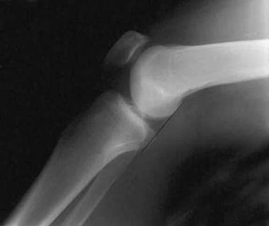
See the image below.
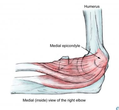 Medial epicondyle.
The ulnar collateral ligament plays a very important role in the surgical treatment for medial epicondylitis and is composed of 3 parts, as follows:
The muscles involved in medial epicondylitis primarily include the pronator teres and the flexor carpi radialis. Less likely to be involved are the palmaris longus, the flexor digitorum superficialis, and the flexor carpi ulnaris.
The anterior medial epicondyle is the primary area of involvement with this condition. The pronator teres partially originates from the superoanterior medial epicondyle, but its primary origin is from the MCT. The AOL is an important valgus stabilizer that must be preserved during surgical intervention.[6] The AOL lies on the posterior margin of the MCT; therefore, the MCT should be located and explored with caution to avoid injury to the AOL.[6] The MCT is not a valgus stabilizer and can be removed if the AOL is intact.
The second most commonly involved muscle is the flexor carpi radialis, which also has a primary origin from the MCT, with a small area of origin from the medial epicondyle. The MCT serves as an important surgical landmark for identification of the involved muscles and in the avoidance of the AOL.[6]
It is important to review the anatomic landmark of the ulnar nerve and the medial antebrachial cutaneous nerve before proceeding with any surgical procedure. At the elbow, the ulnar nerve enters the ulnar groove between the medial epicondyle and the olecranon process. The medial antebrachial cutaneous nerve is in the subcutaneous tissue just proximal to the medial epicondyle, where it divides into the anterior and posterior branches. The posterior branch travels directly over the flexor pronator mass to the posterior medial forearm.[12]
The most common symptom that patients report with medial epicondylitis is achy pain over the anterior medial epicondyle, usually during activity, and the patient may describe weakness in the forearm or hand. In addition, radiation of the pain may occur in the shoulder, forearm, or hand. To evaluate and diagnose this condition, the patient should be questioned regarding trauma, medical, and surgical history; medications; sensory symptoms; and duration and frequency of symptoms.
Begin the examination with inspection of the medial aspect of the affected elbow. Proceed with palpation of the medial and lateral epicondyles, olecranon, brachial pulse, olecranon fossa, capitellum, and radial head. Finally, palpate the triceps, biceps, and flexor and extensor muscles. Palpate the ulnar nerve in the ulnar groove while the patient flexes the arm. In some patients, the ulnar nerve will sublux out of the groove medially over the medial epicondyle. Clinical diagnosis of medial epicondylitis should be considered if tenderness to palpation is present over the anterior aspect of the medial epicondyle. Some patients may have tenderness just distal to the medial epicondyle over the flexor-pronator tendinous bands. The affected elbow's range of motion (ROM) should be normal.
The neurologic examination, including motor, sensory, and reflex testing, is very important to exclude cervical radiculopathy and ulnar neuropathy. Evaluate for a Tinel sign between the olecranon and the medial epicondyle to detect ulnar neuropathy (ie, lightly tapping on the nerve exacerbates the patient's symptoms and/or results in a tingling paresthesia). Pain over the medial epicondyle should be worse with resisted wrist flexion and pronation. Valgus and varus stress testing should be performed to evaluate for ulnar and radial collateral ligament instability[13] . Surgery is contraindicated in the presence of any ligamentous instability.
Standard radiography should be performed to evaluate for other pathology, such as trauma and osteoarthritis. If medial instability is suspected, valgus stress radiographs are recommended.[14]
MRI is sensitive and specific in the evaluation of medial epicondylitis. This modality allows assessment of the tendons, ulnar nerve, and medial collateral ligament.[15] However, MRI should be limited to clinically challenging cases.
Ultrasonography may be used to visualize the degenerative tendons that are involved with medial epicondylitis.[15, 16] Ultrasonography has been shown to be sensitive and specific in the diagnosis of medial epicondylitis. In addition, it can help differentiate tendinosis from partial-thickness or intrasubstance tears.[17]
Electromyography is indicated in cases in which ulnar neuropathy may be associated with this condition.
In an injection test, approximately 1 mL of 2% lidocaine can be injected into the medial epicondyle over the area of maximal tenderness. If the patient has complete relief of pain, then a diagnosis of medial epicondylitis can be made.
The mainstay of treatment for medial epicondylitis is conservative management, which includes the following:
The patient should be educated about the condition's contributing factors and activity modification. Increased wrist flexion and pronation should be avoided. If ulnar neuropathy is associated with the medial epicondylitis, the patient should avoid elbow flexion and leaning on the elbow. In addition, in the immediate term, the patient should place ice packs on the medial epicondyle for 10-15 minutes 3-4 times per day to decrease the inflammation that occurs early in medial epicondylitis.
McCarroll reported that most elbow injuries that are seen during the sport of golf occur during impact.[18] The author recommended that golfers with medial epicondylitis should seek a professional instructor for the proper technique and equipment. Golf-swing modification should begin with a smooth back swing, with the wrist cocking naturally. During the back swing–to–impact transition, motion should begin in the hips to reduce stress in the arms and elbow. Also, McCarroll recommended that the forward arm motion should be initiated by the shoulder, not the wrists. The common incorrect swing of casting the club into the swing or hitting from the top can be a major contributor to the development of medial epicondylitis.[18] Moreover, golfers should physically condition themselves to become fit through stretching, strengthening, and cardiovascular exercise before participating in the golf game.
If the patient does not have a contraindication, NSAIDs should be started immediately to decrease inflammation and pain. Be cautious in using these agents in patients who have renal insufficiency, a history of gastrointestinal problems, or both.
Wilks and Andrews developed a rehabilitation protocol to address the acute, subacute, and chronic phases of medial and lateral epicondylitis.[19]
The primary goal of managing the acute phase is to decrease inflammation and pain. Patients are instructed on activity modifications and avoidance of painful movements. Stretching is started to increase flexibility, and therapy modalities such as cryotherapy, whirlpool, phonophoresis, iontophoresis, and friction massage are used with success.
The primary goals of the subacute phase are to improve flexibility, to improve strength, and to increase functional activities. Hand, wrist, elbow, and shoulder strengthening and ROM exercises are started and continued. A counterforce brace/splint is applied. Cryotherapy is used before and after therapy, and the previously painful movements are reinitiated.
The primary goal of the chronic phase is maintenance of strength and flexibility with gradual return to higher-level activities. The flexibility and strengthening exercises are continued, with gradual diminishing of the counterforce brace/wrist splint. Sports activities are gradually reinitiated. The equipment can be modified at this point. Modalities are used in the evening, and a maintenance program/home-exercise program is started.
The wrist splint is placed in a neutral position to rest the flexors. A counterforce brace is applied to the anteromedial/proximal forearm distal to the epicondyle. Avoid placing the medial counterforce brace over the ulnar nerve.[6]
If the patient's symptoms persist after 2 weeks of conservative management, then consider a corticosteroid injection. With a 25-gauge 0.5-inch needle, inject 1-2 mL of equal amounts 2% lidocaine and triamcinolone acetonide (Kenalog) at 10 mg/mL over the medial epicondyle at the area of maximal tenderness. Direct the patient to position the elbow in extension to avoid the ulnar nerve because some patients may have ulnar nerve subluxation with flexion. To avoid the tendon, never inject under pressure; and to avoid the ulnar nerve, do not inject if the patient reports a sharp, radiating sensation.
Stahl and Kaufman conducted a prospective, randomized, double-blind study to analyze the short- and long-term effects of local injections of methylprednisolone for the treatment of medial epicondylitis.[20] Fifty-eight patients were monitored over 1 year. The authors concluded that the local injection provided only short-term (up to 6 wk) benefits. Compared with the group that did not receive steroids, the outcome was the same with regard to pain at 3 months and at 1 year. Therefore, the injection should be considered early in treatment, with the goal of pain relief.
Other techniques, such as low-level laser therapy and shockwave therapy, have been attempted but have been shown to be unsuccessful.[21]
Surgical treatment should be considered in cases in which conservative treatment has failed after 6-12 months and after all other pathology has been excluded. Medial epicondylitis is classified based on the presence and severity of concomitant ulnar neuropathy. Type IA has no associated ulnar neuropathy, type IB has mild symptoms that are associated with ulnar neuropathy, and type II has moderate to severe symptoms that are associated with ulnar neuropathy.[22]
Gabel and Morrey described the following classification system to guide surgical treatment[6] :
The following technique is used for type IA medial epicondylitis.[6] Variations with epicondylar debridement can be used, depending on the literature and surgical preference.
Epicondylar debridement begins with a 3- to 7-cm incision just anterior to the medial epicondyle. The posterior division of the medial antebrachial cutaneous nerve must be identified and avoided during the approach through the subcutaneous tissue. The common flexor pronator origin is identified, and in order to protect the ulnar nerve, the nerve is identified in the ulnar groove. The flexor pronator fascia is incised, and a 2-mm rim of superficial fascia is preserved on the medial epicondyle for later repair. The lesion is identified at the MCT, and the AOL is also identified. The lesion is excised, and the AOL is protected for elbow stability. The anterior cortex is roughened with a curette or by drilling multiple small holes to increase the blood supply. The common flexor pronator origin is then repaired to the superficial fascia with interrupted sutures, and the subcutaneous tissue and skin are closed in routine fashion.
In type IB cases with ulnar nerve compression, the cubital tunnel release is performed along with epicondylar debridement. If ulnar subluxation or adhesions are encountered, a submuscular transposition is performed. In type II cases, a debridement is performed with a submuscular transposition of the ulnar nerve.[6]
Postoperative management includes an immobilizing splint or cast for 1-3 weeks. ROM exercises are started after removal of the splint or cast. A stretching and strengthening flexor-pronator program is started 6 weeks after surgery.
Surgical results correlate with the type of medial epicondylitis. Type IA or IB medial epicondylitis has a 95% good or excellent postoperative success rate.[6] Type II cases that have more involved ulnar neuropathy have a poorer prognosis secondary to the failure of the neuropathy to respond to surgical management.[6]
Medial epicondylitis is not routinely managed arthroscopically. Some authorities report an arthroscopic technique for lateral epicondylitis. A cadaver study suggests that arthroscopic debridement of the medial epicondyle can be performed, with a low potential for injury to the medial collateral ligament or the ulnar nerve.[23]
Kwon et al conducted a retrospective review to evaluate the use of fascial elevation and tendon origin resection (FETOR) for surgical treatment of medial epicondylitis in 22 elbows of 20 patients (mean age, 48.8 years); the mean follow-up period was 35.6 months.[24] Outcome assessments included the visual analogue scale (VAS) for average pain, pain at rest, and pain experienced during hard work or heavy lifting; the Disabilities of the Arm, Shoulder and Hand (DASH) questionnaire; and assessment of pain-free grip strength.
Comparison of preoperative and postoperative data revealed significant improvement with FETOR in all measures used in assessing pain and strength.[24] There were no major complications. The investigators concluded that the FETOR technique is a safe and effective method treating chronic, recalcitrant medial epicondylitis.
Poor prognosticating factors for medial epicondylitis include work activities with high levels of strain, particularly with non-neutral wrist postures.[25]
Recognizing and treating medial epicondylitis in the acute stage is very important. The long-term complications of the untreated condition include chronic pain, loss of function, and possible elbow contracture.[26] Other important complications include the well-known adverse effects of NSAIDs, most commonly gastrointestinal bleeding, ulceration, and renal dysfunction. Local steroid injections may increase the risk of infection, skin pigmentary changes and skin atrophy, ulnar nerve damage, and tendon rupture.[26]
Complications of surgical treatment include restricted ROM, neuropathic pain from injury to the medial antebrachial cutaneous nerve or ulnar nerve, and medial elbow instability. In most patients, medial epicondylitis resolves with conservative treatment at 6 months, but this may take up to 2 years.
Cho et al reported the results of mini-open muscle resection for intractable lateral or medial epicondylitis in 42 elbows following 6 months of conservative treatment, a history of more than 3 steroid injections, or severe functional impairment.[27] Average preoperative VAS scores of pain were 5.36 at rest, 6.44 at daily activities, and 8.2 at sports or occupational activities. After surgery, the VAS scores were 0.3 at rest, 1.46 at daily activities, and 2.21 at sports or occupational activities.
Preoperative Roles and Maudsley scores were acceptable in 6 cases and poor in 36 cases; postoperatively, the scores were excellent in 23 cases, good in 16 cases, and acceptable in 3 cases.[27] Overall, 41 cases (97.6%) achieved satisfactory results, according to the authors. Postoperative complications consisted of 2 cases of subcutaneous seroma due to leakage of joint fluid, which was successfully managed by additional surgery and suction drainage.
Shahid et al conducted a retrospective study to assess outcomes of open surgery for patients with recalcitrant medial epicondylitis following failed conservative management.[28] The investigators reviewed clinical results for 15 patients (17 elbows). The mean follow-up period of the study cohort was 66 months.
In this study, operative treatment improved patient function significantly.[28] There was a mean increase of 10 kg in grip strength and a mean decrease (improvement) of 25.7 in the DASH score. All but 1 patient experienced little or no residual elbow discomfort and had excellent Mayo elbow performance scores postoperatively. Eleven of the 12 patients who were previously employed were able to return to work within 8 weeks of surgery.
Copyright © www.orthopaedics.win Bone Health All Rights Reserved