Shoulder dislocation is the most common large-joint dislocation seen in the emergency department (ED). The muscular, ligamentous, and bony anatomy of the shoulder (glenohumeral joint) gives it the most extensive range of motion of any joint in the human body. However, this anatomy also makes the glenohumeral joint the most unstable joint in the body.
Anterior dislocations (in which the humeral head is displaced anteriorly in relation to the glenoid), account for as many as 95-98% of shoulder dislocations.[1] The reason is that the muscular and ligamentous support anterior to the humeral head is much less robust than the substantial muscular and bony support afforded posteriorly by the rotator cuff and scapula. Anterior shoulder dislocations may be divided into the following four types:
Posterior shoulder dislocations are considerably less common, accounting for no more than 4% of all shoulder dislocations. Perhaps for this reason, many posterior shoulder dislocations are initially missed by treating physicians, and diagnosis is delayed in nearly all cases.[2] Failure to diagnose and treat posterior dislocations promptly can result in complications, including recurrent dislocations, avascular necrosis of the humeral head, degenerative disease, and chronic pain.
Inferior glenohumeral dislocation (luxatio erecta humeri) is rare, accounting for fewer than 1% of all shoulder dislocations.[3, 4] Most cases arise from forceful hyperabduction of the shoulder. This initially results in impingement of the humeral head against the acromion, and the leverage caused by this impingement ultimately drives the humeral head downward, causing it to disrupt the inferior portion of the glenohumeral capsule and dislocate. Forceful, direct axial loading of an abducted shoulder can also result in luxatio erecta.[5, 6]
Most shoulder dislocations are straightforward and can easily be reduced in the ED by using one of several techniques. However, difficult cases do occur, and clinicians need to be alert for coincident injuries and complications.
NextFor subcoracoid and subglenoid dislocations, which account for 99% of anterior shoulder dislocations, joint reduction by the ED physician is typically indicated. Subclavicular or intrathoracic dislocations, which are caused by large forces, are not easily corrected and should be referred to an orthopedic surgeon.[7]
For an uncomplicated posterior shoulder dislocation that is diagnosed within 6 weeks of injury, reduction in the ED is generally appropriate. A small humeral head defect is not a contraindication for attempting a closed reduction in the ED. A fracture-dislocation with a nondisplaced lesser tuberosity fracture may be treated with a closed reduction.
In a patient with an inferior glenohumeral dislocation, the presence of brachial plexus injury necessitates prompt, atraumatic reduction, with the goal being smooth, uncomplicated, successful reduction on the first attempt.
Standard closed reduction of an anterior shoulder dislocation is absolutely contraindicated if prompt surgical consultation is indicated.[7] It is ruled out by the following:
Various neurovascular injuries and common fractures do not prohibit reduction but do call for prompt and atraumatic reduction with avoidance of multiple attempts. These include the following:
The following are contraindications for standard closed reduction of a posterior shoulder dislocation:
Standard closed reduction of an inferior glenohumeral dislocation is contraindicated in the setting of humeral neck or shaft fractures or in the setting of suspected major vascular injury. The presence of these associated injuries necessitates surgical intervention/open reduction.
Though not a contraindication per se, a “buttonhole deformity” (in which the humeral head becomes trapped in a tear of the inferior capsule) often precludes successful closed reduction, necessitating open reduction.
Clinical assessment determines the type of dislocation present, which guides the approach to reduction (if indicated).
Anterior dislocation
A patient with an anterior shoulder dislocation typically presents with an obvious squared-off shoulder, with the humeral head located inferior and medial to the normal anatomic location. Patients generally hold the injured arm in abduction and resist attempts to adduct or internally rotate the arm.[7] Trying to place the arm into a sling is often futile; patients usually find the position of greatest comfort.
Before any attempts at reduction, the provider should perform a neurovascular examination and assess the probability of a fracture, considering the mechanism of injury and the physical characteristics of the patient. The axillary nerve is the most commonly injured nerve in shoulder dislocations and can be evaluated by testing for sensation in the lateral upper arm and by palpating for contraction of the deltoid muscle while the patient abducts against resistance. The clinician should also look for possible damage to other branches of the brachial plexus.
Arterial injury, though rare in this setting, is also possible and can present with paresthesias, diminished pulse, paleness or coolness of the affected extremity, pain that is out of proportion to the physical findings, or paralysis.[7] Injury to the axillary artery is more common in the elderly population.[8]
Posterior dislocation
Posterior shoulder dislocations usually result from forceful contractions of the internal rotators that occur during seizures and electrical shock. This mechanism can force the humeral head posteriorly, out of its normal alignment and behind the glenoid. Less commonly, posterior shoulder dislocations follow trauma. The mechanism may be a direct blow to the anterior shoulder or a posteriorly directed force applied through the forward-flexed arm.
A complete neurovascular examination should be performed for these dislocations as well, though the incidence of neurovascular injuries is lower with posterior dislocations than with anterior dislocations.[9] Posterior shoulder dislocations are commonly associated with posterior glenoid rim fractures and anterior compression fractures of the humeral head.
Inferior dislocation
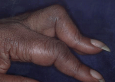
Patients with inferior glenohumeral dislocations present with the affected arm “locked” in abduction of varying degrees.[10] Classically, the affected arm is hyperabducted, with the elbow flexed and the forearm resting on top of or behind the head (see the image below). Often, the dislocated humeral head is palpable along the lateral border of the chest wall. The patient is generally in a substantial amount of pain, particularly when attempts are made to move the injured extremity.
 Classic presentation of inferior shoulder dislocation. Affected arm is hyperabducted, with elbow flexed and forearm resting on top of head.
In some cases of anterior shoulder dislocation, standard reduction efforts will fail. If multiple attempts at closed reduction fail or signs of neurovascular injury develop, an orthopedic surgeon should be consulted to evaluate for closed reduction or possible open reduction in the operating room with general anesthesia.
Injuries may occur as a consequence of reduction. These may be minimized by applying the smallest effective amount of force during reduction with traction and leverage techniques so as to avoid the formation or exacerbation of existing fractures or vascular (eg, hemarthrosis) or nerve injuries (eg, neurapraxia). New fractures rarely appear on postreduction films.
Recurrence is the most common complication adverse outcome after reduction, especially in young active patients. Age at the time of dislocation is inversely related to the rate of recurrence.[11] Common fractures (eg, Hill-Sachs deformity or Bankart fracture) require prompt orthopedic follow-up because they are associated with increased joint instability and a higher risk of redislocation. After evaluation of the shoulder’s postreduction range of motion, immediate immobilization with a sling and swathe is crucial to prevent recurrence.
The most common complication of attempted closed reduction of a posterior shoulder dislocation is a humeral fracture. Acute redislocation may also occur. Complications of the dislocation itself include the following:
An estimated 50-60% of patients with luxatio erecta have associated brachial plexus injury.[12] Assessment and documentation of the presence of neurologic deficits should be carried out both before and after reduction.[13] Injury to the axillary artery, including arterial thrombosis, has also been reported.[14]
Rotator cuff tears occur very often with inferior dislocations.[15, 16] Ligamentous and connective tissue injuries include disruption of the glenohumeral ligament, the inferior glenoid capsule, or both.
Associated bony injuries include fractures of the glenoid rim, greater tuberosity, acromion, clavicle, and coracoid process.[2] These injuries can be induced or exacerbated by attempted reduction; however, they more often occur as a result of the dislocation itself.
Periprocedural Care
Copyright © www.orthopaedics.win Bone Health All Rights Reserved