Osteomyelitis is inflammation of the bone caused by an infecting organism. Although bone is normally resistant to bacterial colonization, events such as trauma, surgery, the presence of foreign bodies, or the placement of prostheses may disrupt bony integrity and lead to the onset of bone infection. Osteomyelitis can also result from hematogenous spread after bacteremia. When prosthetic joints are associated with infection, microorganisms typically grow in biofilm, which protects bacteria from antimicrobial treatment and the host immune response.
Early and specific treatment is important in osteomyelitis, and identification of the causative microorganisms is essential for antibiotic therapy.[1] The major cause of bone infections is Staphylococcus aureus. Infections with an open fracture or associated with joint prostheses and trauma often must be treated with a combination of antimicrobial agents and surgery. When biofilm microorganisms are involved, as in joint prostheses, a combination of rifampin with other antibiotics might be necessary for treatment.
NextThe bony skeleton is divided into two parts: the axial skeleton and the appendicular skeleton. The axial skeleton is the central core unit, consisting of the skull, vertebrae, ribs, and sternum; the appendicular skeleton comprises the bones of the extremities. The human skeleton consists of 213 bones, of which 126 are part of the appendicular skeleton, 74 are part of the axial skeleton, and six are part of the auditory ossicles.
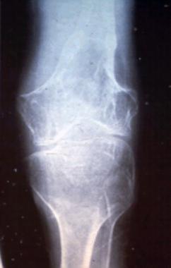
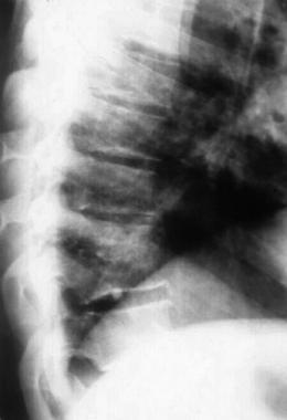
Hematogenous osteomyelitis most commonly involves the vertebrae, but infection may also occur in the metaphysis of the long bones, pelvis, and clavicle. Vertebral osteomyelitis involves two adjacent vertebrae with the corresponding intervertebral disk. (See the image below.) The lumbar spine is most commonly affected, followed by the thoracic and cervical regions.
 Osteomyelitis of T10 secondary to streptococcal disease. Photography by David Effron MD, FACEP.
Posttraumatic osteomyelitis begins outside the bony cortex and works its way in toward the medullary canal, typically found in the tibia. Contiguous-focus osteomyelitis often occurs in the bones of the feet in patients with diabetes mellitus and vascular compromise.
For more information about the relevant anatomy, see Skeletal System Anatomy in Adults and Osteology (Bone Anatomy).
Bone is normally resistant to infection. However, when microorganisms are introduced into bone hematogenously from surrounding structures or from direct inoculation related to surgery or trauma, osteomyelitis can occur. Bone infection may result from the treatment of trauma, which allows pathogens to enter bone and proliferate in the traumatized tissue. When bone infection persists for months, the resulting infection is referred to as chronic osteomyelitis and may be polymicrobial. Although all bones are subject to infection, the lower extremity is most commonly involved.[1, 2]
Some important factors in the pathogenesis of osteomyelitis include the virulence of the infecting organism, underlying disease, immune status of the host, and the type, location, and vascularity of the bone. Bacteria may possess various factors that may contribute to the development of osteomyelitis. For example, factors promoted by S aureus may promote bacterial adherence, resistance to host defense mechanism, and proteolytic activity.[3]
In adults, the vertebrae are the most common site of hematogenous osteomyelitis, but infection may also occur in the long bones, pelvis, and clavicle.[4]
Primary hematogenous osteomyelitis is more common in infants and children, usually occurring in the long bone metaphysis. However, it may spread to the medullary canal or into the joint. When infection extends into soft tissue, sinus tracts may eventually form. Secondary hematogenous osteomyelitis is more common and occurs when a childhood infection is reactivated. In adults, the location is also usually metaphyseal.[4]
S aureus is the most common pathogenic organism recovered from bone, followed by Pseudomonas and Enterobacteriaceae. Less common organisms involved include anaerobe gram-negative bacilli. Intravenous drug users may acquire pseudomonal infections. Gastrointestinal or genitourinary infections may lead to osteomyelitis involving gram-negative organisms. Dental extraction has been associated with viridans streptococcal infections. In adults, infections often recur and usually present with minimal constitutional symptoms and pain. Acutely, patients may present with fever, chills, swelling, and erythema over the affected area.[2, 5]
The initiating factor in contiguous-focus osteomyelitis often consists of direct inoculation of bacteria via trauma, surgical reduction and internal fixation of fractures, prosthetic devices, spread from soft-tissue infection, spread from adjacent septic arthritis, or nosocomial contamination. Infection usually results approximately one month after inoculation.
Posttraumatic osteomyelitis more commonly affects adults and typically occurs in the tibia. The most commonly isolated organism is S aureus. At the same time, local soft-tissue vascularity may be compromised, leading to interference with healing. Compared with hematogenous infection, posttraumatic infection begins outside the bony cortex and works its way in toward the medullary canal. Low-grade fever, drainage, and pain may be present. Loss of bone stability, necrosis, and soft tissue damage may lead to a greater risk of recurrence.[4, 5]
Septic arthritis may lead to osteomyelitis. Abnormalities at the joint margins or centrally, which may arise from overgrowth and hypertrophy of the synovial pannus and granulation tissue, may eventually extend into the underlying bone, leading to erosions and osteomyelitis. One study demonstrated that septic arthritis in elderly persons most commonly involves the knee and that, despite most of the patients having a history of surgery, 38% developed osteomyelitis. Septic arthritis is more common in neonates than in older children and is often associated with metaphyseal osteomyelitis. Although rare, gonococcal osteomyelitis may arise in a bone adjacent to a chronically infected joint.[6, 7]
Patients with vascular compromise, as in diabetes mellitus, are predisposed to osteomyelitis owing to an inadequate local tissue response.[4]
Infection is most often caused by minor trauma to the feet with multiple organisms isolated from bone, including Streptococcus species, Enterococcus species, coagulase-positive and -negative staphylococci, gram-negative bacilli, and anaerobic organisms. Foot ulcers allow bacteria to reach the bone. Patients may not experience any resulting pain, because of peripheral neuropathy, and may present with a perforating foot ulcer, cellulitis, or an ingrown toenail.
Physical examination may reveal decreased sensation, poor capillary refill, and decreased dorsalis pedis and posterior tibial pulses. Treatment is aimed at suppressing infection and improving vascularity. However, most patients develop recurrent or new bone infections. Resection or amputation of the affected tissue is sometimes necessary. Debridement, incision and drainage, and tendon lengthening are attempted first.
The incidence of vertebral osteomyelitis generally increases progressively with age, with most affected patients being older than 50 years. Although devastating complications may result from a delay in diagnosis, vertebral osteomyelitis is rarely fatal since the development of antibiotics. The infection usually originates hematogenously and involves two adjacent vertebrae with the corresponding intervertebral disk. The lumbar spine is most commonly affected, followed by the thoracic and cervical regions.[4, 1]
Potential sources of infection include skin, soft tissue, respiratory tract, genitourinary tract, infected intravenous sites, and dental infections. S aureus is the most common isolated organism. However, Pseudomonas aeruginosa is more common in intravenous drug users.
Most patients with vertebral osteomyelitis present with localized pain and tenderness of the involved vertebrae with a slow progression over 3 weeks to 3 months. Fever may be present in approximately 50% of patients. Fifteen percent of patients may have motor and sensory deficits. Laboratory studies may reveal peripheral leukocytosis and an elevated erythrocyte sedimentation rate. Extension of the infection may lead to abscess formation.[4]
Acute hematogenous osteomyelitis usually occurs after an episode of bacteremia in which the organisms inoculate the bone. The most common organisms isolated in these cases include S aureus, Streptococcus pneumoniae, and Haemophilus influenza type b (less common since the use of vaccine for H influenza type b).
Acute hematogenous S aureus osteomyelitis in children can lead to pathologic fractures. This can occur in about 5% of cases with a 72-day mean time from disease onset to fracture.[8]
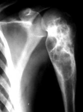
In children with subacute focal osteomyelitis (see the image below), S aureus is the most commonly isolated organism.
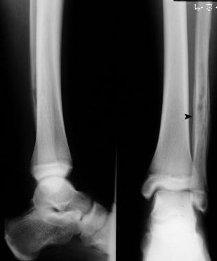 Rarefaction and periosteal new-bone formation around the left upper fibula in a 12-year-old patient. This was caused by subacute osteomyelitis.
Gram-negative bacteria such as Pseudomonas species or Escherichia coli are common causes of infection after puncture wounds of the feet or open injuries to bone. Anaerobes can also cause bone infection after human or animal bites.
Osteomyelitis in the neonate results from hematogenous spread, especially in patients with indwelling central venous catheters. The common organisms in osteomyelitis of the neonate include those that frequently cause neonatal sepsis, namely group B Streptococcus species, and E coli. Infections in the neonate can involve multiple osseous sites, and approximately half of the cases also involve eventual development of septic arthritis in the adjacent joint.
Children with sickle cell disease are at an increased risk for bacterial infections, and osteomyelitis is the second most common infection in these patients. The most common organisms involved in osteomyelitis in children with sickle cell anemia include Salmonella species, S aureus, Serratia species, and Proteus mirabilis.
Posttraumatic osteomyelitis accounts for as many as 47% of cases of osteomyelitis. Other major causes of osteomyelitis include vascular insufficiency (mostly occurring in persons with diabetes; 34%) and hematogenous seeding (19%).
Motor vehicle accidents, sports injuries, and the use of orthopedic hardware to manage trauma also contribute to the apparent increase in prevalence of posttraumatic osteomyelitis. Osteomyelitis may complicate puncture wounds of the foot, occurring in 1.8%-6.4% of patients following injury.[9, 10, 11, 12, 13]
Approximately 20% of adult cases of osteomyelitis are hematogenous, which is more common in males for unknown reasons.[4]
The incidence of spinal osteomyelitis was estimated to be 1 in 450,000 in 2001. In subsequent years, however, the overall incidence of vertebral osteomyelitis is believed to have increased as a consequence of intravenous drug use, increasing age of the population, and higher rates of nosocomial infection due to intravascular devices and other instrumentation.[14, 15] The overall incidence of osteomyelitis is higher in developing countries.
Inadequate therapy may lead to relapsing infection and progression to chronic infection. Because of the avascularity of bone, chronic osteomyelitis is curable only with radical resection or amputation. These chronic infections may recur as acute exacerbations, which can be suppressed by debridement followed by parenteral and oral antimicrobial therapy. Rare complications of bone infection include pathologic fractures, secondary amyloidosis, and squamous cell carcinoma at the sinus tract cutaneous orifice.
Clinical Presentation
Copyright © www.orthopaedics.win Bone Health All Rights Reserved