QuestionQUESTION: Straight to the point I shall copy my results.
MRI Lumbar Spine: Sagittal and axial Mr Images. Satisfactory vertebral alignment. No fracture or focal bone lesion.
The upper three lumbar intervertebral discs are normal. At L4/5 is loss of disk hydration and subtle disc space narrowing. A small posterior disc extrusion extends posteriorly and inferiorly deep to the posterior longitudinal ligament. This disc material indents the thecal sac, resulting in minor central canal stenosis. Mild bilateral foraminal narrowing also noted, however, no convincing evidence of focal nerve root compression. The L5/S1 disc is normal.
No significant facet joint arthropathy. Paravertebral soft tissues are normal.
MRI right Hip: Degenerative changes are present at the right hip with mild diffuse chondral thinning on both sides of the joint and mild joint space narrowing. Osteophytic lipping is prominent along the superolateral aspect of the femoral head. There is a small round area of fluid signal change at the base of the fibrocartilaginous labram consistent with labral degeneration, however, I do not identify a discrete labral tear. A small joint effusion is present. No intraarticular loose body.There is mild tendinopathy involving the anterior fibres of the distal gluteus medius tendon. A thin layer of fluid is present in the trochanteric bursa. Remaining regional musculature normal.
Any help appreciated I am 43 years old, have had long term iron deficency requiring course of injections every now and then. I have been diagnosed with glucose intolerance but a recent blood test showed ok levels although it was not a fasting test. I experience overloads of yeast occasionaly as well in the throat and intestines, the pain will vary day to day. I am in pain most nights and dont sleep well. My legs at times can be weak. I get sore from either moving or rest depends on day and sometimes walk to ease other times I have to rest to ease.
Some people dont seem to understand the pain I am in at times and I get teased about just getting old. Any suggestions would be appreciated in pain management and further treatment
ANSWER: Dear Shaz,
Your MRI results have some common degenerative changes seen in patients with and without low back pain and hip pain. The areas of concern are at L4/L5 due to the fact that you have bulging disc material which is pressing on the thecal sac (covering of the spinal cord) The thecal sac has a rich innervation of pain fibers which can account for low back symptoms. Stenosis means that the area around the spinal cord has been made smaller which can result in local inflammation to the spinal cord and associated structures such as the nerve roots that travel down the legs. However, the repot does not find that the nerve roots have been compressed which is good.
The right hip joint is degenerative with some bony overgrowth (arthritic changes), and the cartilage that lines the hip joint (labrum) has degeneration as well. Again this can easily account for localized pain around the right hip, and this is complicated by the fact that you have some tendinitis (inflammation of the end of the muscles) in the butt muscles that attach around the hip joint.
With the above in mind, yes you have degenerative conditions in the low back disk as well as the right hip, but have you been given any opinion why the degeneration started in the first place? The reason I ask this is because i often see degeneration in my patient who do not have pain, and pain in patients without degeneration. So your doctors need to get to the root cause. I would take an educated guess that you have had prolonged hip and/or spinal dysfunction for years that has gone un-noticed, and that it needs to be corrected first. This can often be as simple as correcting the joint mechanics with exercise and stretching protocols, however it can also be more complicated that that requiring specific traction protocols as well. Not to mention that the soft tissue of the gluteus musculature needs to be addressed to lessen the tendinitis in that area...often deep tissue work will be needed such as Active release techniques to free up the tissue and break up scar formation. Bottom line here is that you need a multidisciplinary functional care approach such as myofascial release, chiropractic care, functional physical therapy, and possible structural rehab of the spine. I would bet the structure of your spine and sacrum are out of line...according to the published normative data. (published in the journal, SPINE)
Some suggestions: try to find a good chiropractor that is trained in sports injury, active release, graston or chiropractic biophysics in your area. If you can find one that has 2 of these certifications, that is even better. Why?...because these chiropractors will apply active care protocols for rehabilitation not just give adjustments and send you out the door. Active care protocols have been shown to procure better results in pain reduction and increased function.
www.acbsp.com
www.idealspine.com
www.activerelease.com
www.grastontechnique.com
Considering the yeast (candida) issue, this is a common problem, but one that is often not well managed by traditional medical protocols. The reason is because yeast is resistant to pharmacological management, and often the only approach taken in prescription medication of Diflucan (FLUCONAZOLE). It is common knowledge in medical circles that Diflucan is only marginally effective because yeast has become resistant to it. This is compounded by your glucose issues, and if you live in the southern region of the country, the climate will exacerbate this as well.
Amazingly though, newer research from dentists, medical docotrs, chiropractors, nutritionists, and biochemistry professionals has found alternative techniques to manage and eliminate the problem...and numerous good books have been written on the topic of yeast overgrowth. I would suggest that you seek out a few books on this topic (books with multiple scientific references only) and read them...you may be able to find some at the local library. Organic foods, probiotic supplements, avoidance of refined carbohydrates, and sugar will all help to reduce yeast in the body. Gentian violet is another substance that will quickly eliminate yeast, however it is to be used more topically than internally. This is utilized mostly with breast feeding mothers who get candida infections of the breast and for the infants who get the infections in their mouths. It can be utilzed orally, but in small dosages only.
Hope this helps.
Respectfully,
Dr. J. Shawn Leatherman
www.suncoasthealthcare.net
---------- FOLLOW-UP ----------
QUESTION: Thank you, your assistance has been very valuable.
I am seeing an osteopath and am sourcing a pilates class close to home. Unfortunately I work long extreme hours and don't put enough aside to take car of my self.
I am wearing a heel raiser as it makes me feel like I am walking normal and without it for a day or more will inflame the area, my osteopath suggested this due to the droop of my hip. He believes I could have been walking around with one leg shorter than the other for some time.
My question is could it all be connected with my iron deficiency as this has never been discovered as to why.(Appointment coming up with gastro Dr in october.)
I was in hospital at 4yrs for a week undergoing tests as I was unwell, nothing was found and it was put down to being anemic. I was wondering could this affect the muscles as it was only really discovered how low in iron I was when I was 39 and no supplements were ever taken until then, could it have compromised my growth or muscle development etc as my muscles have always been tender to touch with some pressure particularly my legs. I don't hear other people complain of similar symptoms as my self and I know my tolerance to pain is quite high enduring 4 natural births. I will put more effort into stretching etc to help myself but I cant help thinking there is an underlying syndrome there undetected.
Thank you again
Shaz
AnswerDear Shaz,
Having a leg length inequality is actually a fairly common finding in patients, however, most doctors never actually look for the relationship of a short leg to spinal or pelvic dysfunction. I am actually impressed that your Osteopath has analyzed the relationship and given you a heel lift to correct the structural differences.
In regards to your previous question about the MRI, leg length inequality can lead to accelerated degeneration in the lumbar spine and sacroiliac joints of the pelvis due to long term altered weight bearing on the joints. So this can easily be a causative factor in the degeneration found, however, it is probably not the sole factor. I would definitely recommend that you keep wearing the heel lift, but I would also recommend that you have follow-up AP x-ray of the pelvis and lumbar spine to see if the heel lift you are utilizing correctly balances the structures. A simple one shot x-ray view while standing in your shoes with the lift in place. You can under or over correct a short leg with a heel lift, and the lift needs to maximize leg length correction without significantly tilting the sacrum to one side, or leaning the spine off to one side. I have sometimes found that the size of the heel lift to be utilized does not match the measurements on the x-ray film...that is why it is nice to rake a follow-up view a few weeks after we have placed it.
Now, I doubt that your iron deficiency anemia has anything to do with the differences. Leg length inequality happens all the time, and the proposed factors of why it happens are varied. Theories are from fetal development, altered weight bearing in childhood such as wearing backpacks on one shoulder only, minor childhood injuries, vigorous contact sports before skeletal maturity etc... Nutritional deficiency has been implicated as well, however this would result in a systemic or body wide condition such as rickets in children where the deformity is on both sides of the body...not just one. So again I doubt there is a relationship. Other than a frank injury resulting in a fractured long bone or growth plate in the leg, it really is speculative.
However, when you are anemic, the red blood cells in the body have a decreased capacity to deliver oxygen to the tissues which may have the effect of not allowing the muscular system an adequate oxygen supply in times of physical exertion. Everyone know what it feels like to have sore muscle after a workout due to the build-up of muscle breakdown products in the blood...lactic acid predominately, but also some other chemical mediators of pain. What people do not realize is that this mechanism is mainly caused by a lack of oxygenation to the tissue from depletion...one of the reasons we breathe harder when we exercise. Anyway, if you are anemic-low red cell counts, you have less red blood cell volume to get oxygen to the tissues, and this could be one factor in the increased muscle soreness.
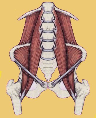
Moving on, referred pain from the SI joint due to the leg length inequality, from the degenerative joints in the low back, or degenerative disks can also create chronic leg pain. This is called sclerotogenous pain referral. I am going to attach a few documents to the end of this response for you to appreciate on pain generated from the tissue of the pelvis and low back...they are a synopsis of current research papers and may be difficult to fully understand, but you will get the point and it should broaden your understanding of possible pain mechanisms. Hope this helps Shaz.
Respectfully,
Dr. J. Shawn Leatherman
www.suncoasthealthcare.net
The Role of Sacroiliac Joint Dysfunction in the Genesis of Low Back Pain: The obvious is not always right
Archives of Orthopaedic and Trauma Surgery
December 2007 [e-pub]
Natan Weksler, Gad J. Velan, Michael Semionov, Boris Gurevitch, Moti Klein, Vsevolod Rozentsveig and Tzvia Rudich
FROM ABSTRACT:
Background context: It is a common practice to the link low back pain with protruding disc even when neurological signs are absent. Because pain caused by sacroiliac joint dysfunction can mimic discogenic or radicular low back pain, we assumed that the diagnosis of sacroiliac joint dysfunction is frequently overlooked.
Purpose: To assess the incidence of sacroiliac joint dysfunction in patients with low back pain and positive disc findings on CT scan or MRI, but without claudication or objective neurological deficits.
Methods: Fifty patients with low back pain and disc herniation, without claudication or neurological abnormalities such as decreased motor strength, sensory alterations or sphincter incontinence and with positive pain provocation tests for sacroiliac joint dysfunction were submitted to fluoroscopic diagnostic sacroiliac joint infiltration.
Results: The mean baseline VAS pain score was 7.8. Thirty minutes after infiltration [of the SI joint with anesthesia], the mean VAS score was 1.3. Forty-six patients [out of 50] had a VAS score ranging from 0 to 3, eight weeks after the fluoroscopic guided infiltration.
Conclusions: Sacroiliac joint dysfunction should be considered strongly in the differential diagnosis of low back pain in this group of patients.
THESE AUTHORS ALSO NOTE:
揕ow back pain is second only to common cold as a cause of primary care office visits in the USA.?
揂pproximately 90% of adults have experienced back pain at some point of time in their lives.?br>
Low back pain is responsible for direct care expenditures ranging from $5 billion to more than $20 billion annually and as much as $50 billion per year in indirect costs. Medical residents are not usually taught to consider sacroiliac joint dysfunction as a cause for low back pain.
This study used 200 patients with low back pain who were prior treatment failures from NSAID drugs, physical therapy and intramuscular injections. They were fluoroscopic guided injected with a combination of anesthetic and steroid into their SI joints.
DISCUSSION:
揅hronic persistent low back pain is commonly linked with positive disc findings on CT or MRI imaging. However, these imaging techniques are not always helpful, because they have a poor degree of correlation with clinical signs.? 揑t is not rare to have positive disc findings in asymptomatic patients.?br>
揘early 25% of asymptomatic individuals below the age of 60 years and 33% of older patients have evidence of disc herniation on MRI scans? 揟he diagnosis accuracy of the tissue origin of chronic low back pain and referred lower extremity symptoms based on clinical criteria are about 19?4%.?[In medical practice] Estimated prevalence of SI joints causing low back pain is 13 to 30%.
揟he SIJ has a rich nociceptive innervation.?Its anterior portion is innervated by the posterior rami of the L2朣2 roots, and its posterior aspect is innervated by the posterior rami of L4朣3. [IMPORTANT]
The piriformis muscle is located close to the SIJ, originating at the anterior aspect of the sacrum and inserting into the greater trochanter of the femur. SIJ problems can cause piriformis spasm and provoke sciatic irritation, with 損ain radiating to the buttock, the posterior calf, and to the anterior and lateral calf and foot mimicking radiculopathy.?[IMPORTANT]
揟he current gold standard for the diagnosis of the SIJ syndrome is fluoroscopically guided infiltration of local anesthesia leading to at least an 80% reduction in VAS scores.?However, three physical tests showed good correlation with the diagnosis of SIJ dysfunction: SIJ compression test (94% agreement), the thigh thrust test (90%) agreement, Yeoman抯 test (88%) agreement. [IMPORTANT]
Studies have 揷oncluded that manipulations appear to be successful in many patients suffering from SIJ dysfunction?
CONCLUSIONS:
1) The incidence of SIJ dysfunction in patients with low back pain and discopathy on CT or MRI scans and without neurological deficits appears to be higher than previously described.
2) Pain in SIJ dysfunction can radiate towards the calf and foot mimicking radicular pain.
3) 揚hysicians seeing patients with low back pain should have a high index of suspicion for SIJ dysfunction, especially in the absence of neurological deficits.?
KEY POINTS FROM SUNCOAST HEALTHCARE PROFESSIONALS
1) Because pain caused by sacroiliac joint dysfunction can mimic discogenic or radicular low back pain, the diagnosis of sacroiliac joint dysfunction is frequently overlooked. However, chiropractic physicians are specifically trained to evaluate SIJ function along with lumbar spine function. [IMPORTANT]
2) 揕ow back pain is second only to common cold as a cause of primary care office visits in the USA.?
3) 揂pproximately 90% of adults have experienced back pain at some point of time in their lives.?br>
4) Medical residents are not usually taught to consider sacroiliac joint dysfunction as a cause for low back pain. [IMPORTANT]
5) 揅hronic persistent low back pain is commonly linked with positive disc findings on CT or MRI imaging. However, these imaging techniques are not always helpful, because they have a poor degree of correlation with clinical signs.?br>
6) 揘early 25% of asymptomatic individuals below the age of 60 years and 33% of older patients have evidence of disc herniation on MRI scans?br>
7) Estimated prevalence of SI joints causing low back pain is 13 to 30%.
8) The SI joint has a rich nociceptive innervation by the posterior rami of the L2朣3 roots. [IMPORTANT]
9) The piriformis muscle is located close to the SIJ, originating at the anterior aspect of the sacrum and inserting into the greater trochanter of the femur. SIJ problems can cause piriformis spasm and provoke sciatic irritation, with 損ain radiating to the buttock, the posterior calf, and to the anterior and lateral calf and foot mimicking radiculopathy.?[IMPORTANT]
10) 揟he current gold standard for the diagnosis of the SIJ syndrome is fluoroscopically guided infiltration of local anesthesia leading to at least an 80% reduction in VAS scores.?
11) Three physical tests showed good correlation with the diagnosis of SIJ dysfunction: SIJ compression test (94% agreement), the thigh thrust test (90%) agreement, Yeoman抯 test (88%) agreement. [IMPORTANT]
12) Manipulation is successful in many patients suffering from SIJ dysfunction. [IMPORTANT]
13) The incidence of SIJ dysfunction in patients with low back pain and discopathy on CT or MRI scans and without neurological deficits appears to be higher than previously described.
14) Pain in SIJ dysfunction can radiate towards the calf and foot mimicking radicular pain. [IMPORTANT]
15) 揚hysicians seeing patients with low back pain should have a high index of suspicion for SIJ dysfunction, especially in the absence of neurological deficits.?br>
16) Sacroiliac joint dysfunction should be considered strongly in the differential diagnosis of low back pain in this group of patients.
The nerve supply of the lumbar intervertebral disc
Journal of Bone and Joint Surgery -British Volume, Vol. 89-B, Issue 9, September 2007, pp. 1135-1139
M. A. Edgar, MChir, FRCS, Retired Orthopedic Surgeon
FROM ABSTRACT:
The anatomical studies, basic to our understanding of lumbar spine innervation through the sinu-vertebral nerves, are reviewed.
1980s Research suggested that pain sensation was conducted in part via the sympathetic system. These pathways have now been clarified using sophisticated experimental and histochemical techniques confirming a dual pattern. One route enters the adjacent dorsal root segmentally, whereas the other supply is non-segmental ascending through the paravertebral sympathetic chain with re-entry through the thoracolumbar white rami communicantes.
Sensory nerve endings in the degenerative lumbar disc penetrate deep into the disrupted nucleus pulposus, they do not in the normal lumbar spine. Complex as well as free nerve endings appear to contribute to pain transmission.
The nature and mechanism of discogenic pain is still speculative but there is growing evidence to support a 憊isceral pain?hypothesis, unique in the musculoskeletal system. This mechanism is open to 憄eripheral sensitization?and possibly 慶entral sensitization?as a potential cause of chronic back pain.
THIS AUTHOR ALSO NOTES:
The sinuvertebral nerve is 揻ormed by a fine sympathetic branch, usually arising from the grey ramus communicans, and a fine sensory spinal branch from the ventral ramus.?揟hese conjoined sinuvertebral nerves re-entered the vertebral canal through each intervertebral foramen to lie anterior to the nerve root in association with the segmental vessels.? 揟he sympathetic fibers were considered as vasomotor efferents and the sensory fibers as proprioceptive and nociceptive.?Sinuvertebral nerve branches innervate the posterior longitudinal ligament, the outer layers of the annulus fibrosus, and the anterior dura. 揟he lumbar sinuvertebral nerves had up to three segmental levels of overlap, which might explain the poor localization of low back pain.?br>
The anterior part of the disc annulus is innervated solely from sympathetic nerves. Other parts of the disc are 90% innervated by sympathetic nerves. It is probable that these sympathetic nerves are conveying pain afferent information to the central nervous system, indicating, 搇ow back pain is a kind of visceral pain.?br>
Studies indicate, 揳fferent nerve fibers from the annulus pass into the sympathetic chain to re-enter the sensory nerve roots at L1 and L2.? Other studies state 搕his experiment has confirmed the presence of a clear nociceptive pathway of sympathetic afferent discharge from the dorsal aspect of the lower lumbar intervertebral discs to the dorsal roots of L2,?indicating that 搇umbar discogenic pain is indeed a variety of visceral pain.?
Electrical stimulation to the annulus of an upper lumbar disc produces multilevel bilateral motor unit action potentials in the lumbar multifidus musculature. 揑t is reasonable to propose that the annular stimulus was transmitted via the widespread non-segmental sympathetic afferents.?The pattern of response suggests a spinal reflex to the anterior horn cells.
Electrical stimulus to an adjacent facet joint caused only a localized, unilateral response multifidus contraction. 揇istension of the adjacent facet joint with saline depressed the motor unit action potentials.?[Important for chiropractors-spinal adjustments distend the facet and facet capsule卪ay reduce multifidus spasm]
揘ormal nucleus pulposus and inner annular zones are devoid of nerves.?The three outer lamellae of the disc are innervated with nociceptive afferents. However, nerves can extend to the inner third in 50% of painful degenerative discs. These nerves arise from granulation tissue growing into the degenerative disc, 搉eo-innervation.?Disc and/or facet inflammation can sensitize local mechanoreceptors into becoming pain afferents, resulting in chronic discogenic pain. [Important]
揟he authors of a number of recent papers suggest that the sensory nerve supply of the disc is similar to that of certain enteric structures and represents a form of visceral pain. 揟here is growing evidence that these pain receptors are peripherally sensitized by the activity of sympathetic efferents.?br>
Disc nociceptive afferents 搈ay initiate a pain impulse in response to ischemia, pressure changes (mechanoreceptors) or inflammatory irritation.? [Important for Chiropractors, Vertebral fixation can cause pressure changes affecting mechanoreceptors, while spinal joint cavitation increase mechanoreception from the joint capsules and muscle spindles of the multifidus which inhibits nociception.]
Psychological stress can activate the 慶entral sensitization?of the descending autonomic nerves which may lower the threshold for disc nociception, adding to chronic discogenic pain.
揟here is something unique about the nerves related to the spine and the spinal canal, which makes the source of pain different from the rest of the musculoskeletal parts of the body. Could the answer be that the disc, unlike other joints, is uniquely provided with a predominantly visceral-type of nerve supply??[May be why disc pain is often chronic and requires more treatment than does pain arising from other musculoskeletal tissues]
KEY POINTS FROM SUNCOAST HEALTHARE PROFESSIONALS
1) sympathetic nerves innervate Viscera.
2) The intervertebral disc is also innervated primarily (90%) by sympathetic nerves.
3) Disc sympathetic nerves are capable of sending nociceptive information to the sympathetic nervous system.
4) Disc pain is different than all other musculoskeletal pain because it is a form of 搗isceral pain.?br>
5) The sympathetic component of disc innervation is found in the sinuvertebral nerve. The lumbar sinuvertebral nerves have up to three segmental levels of overlap, 搘hich might explain the poor localization of low back pain.?br>
6) Degenerated discs are more extensively innervated than normal discs.
7) Sensory nerve endings in the degenerative lumbar disc penetrate deep into the disrupted nucleus pulposus, which is insensitive in the normal lumbar spine.
8) The sympathetic nerve fibers that innervate the lower lumbar discs inter the central nervous system through the sensory nerve roots at L1 and L2.
9) 揕umbar discogenic pain is indeed a variety of visceral pain.?
10) Disc irritation produces bilateral contraction of the lumbar multifidus.
11) Facet joint irritation produces unilateral multifidus contraction.
12) Distension of the facet joint inhibits the multifidus muscle contraction. [Important for chiropractors]
13) Disc and/or facet inflammation can sensitize local mechanoreceptors into becoming pain afferents, resulting in chronic discogenic pain. [Important]
14) Pain receptors are sensitized by the activity of sympathetic efferents.
15) Disc nociceptive afferents 搈ay initiate a pain impulse in response to ischemia, pressure changes (mechanoreceptors) or inflammatory irritation.擺Important for Chiropractors]
16) Psychological stress can activate the 慶entral sensitization?of the descending autonomic nerves which may lower the threshold for disc nociception, adding to chronic discogenic pain.
17) 揟here is something unique about the nerves related to the spine and the spinal canal which makes the source of pain different from the rest of the musculoskeletal parts of the body. Could the answer be that the disc, unlike other joints, is uniquely provided with a predominantly visceral-type of nerve supply??br>
Nerve Ingrowth Into Diseased Intervertebral Disc in Chronic Back Pain
The Lancet: July 19, 1997, Vol. 350, pp. 178-181
AJ Freemont, TE Peacock, P Goupille, JA Hoyland, J O払rien, MIV Jayson: Authors associated with the Departments of Rheumatology and Pathological Sciences, University of Manchester, UK.
FROM ABSTRACT:
BACKGROUND:
In the healthy back only the outer third of the annulus fibrosus of the intervertebral disc is innervated.
Nerve ingrowth deeper into diseased intervertebral disc has been reported, but how common this feature is and whether it is associated with chronic pain are unknown. We examined nerve growth into the intervertebral disc in the pathogenesis of chronic low back pain.
METHODS:
We collected 46 samples of intervertebral discs from 38 patients during spinal fusion for chronic back pain. 30 samples were from pain levels clinically established by discography and 16 samples were from adjacent vertebral levels with no pain. We obtained 34 control samples of intervertebral disc from previously healthy individuals with normal histology within 8 h of recorded death. We used standard immunohistochemical techniques to test for a general nerve marker, a nociceptive neurotransmitter (substance P), and a protein expressed during axonogenesis (growth-associated protein 43 [GAP43]).
FINDINGS:
We identified nerve fibers in the outer third of the annulus fibrosus in 48 (60%) of the 80 samples of intervertebral discs. Nerves were restricted to the outer or middle third of the annulus fibrosus in the 34 control samples. Among the patients with chronic low back pain, nerves extended into the inner third of the annulus fibrosus in 46% and into the nucleus pulposus in 22% of samples.
Deep nerve ingrowth into the inner third of the annulus fibrosus, the nucleus pulposus, or both was seen in four (25%) of 16 biopsy samples from non-pain levels and in 17 (57%) samples from pain levels.
Of the 16 paired samples from both pain and non-pain levels, five pain-level samples and one non-pain-level sample showed deep nerve ingrowth.
INTERPRETATION:
Our finding of isolated nerve fibers that express substance P deep within diseased intervertebral discs and their association with pain suggests an important role for nerve growth into the intervertebral disc in the pathogenesis of chronic low back pain.
THESE AUTHORS ALSO NOTE:
These authors cite 5 studies that indicate the intervertebral disc is often the source of back pain and, 揷hronic back pain is one of the leading causes of morbidity and loss of work.?br>
The pathogenesis of chronic back pain is poorly understood, and chronic low back pain is not caused by nerve root compression. Moreover, leg pain / sciatica can be caused by mechanisms other than nerve root compression.
Immunohistochemical staining techniques show that in healthy human intervertebral discs nerves extend no deeper than the outer third of the annulus fibrosis. These authors 搈easured nerve ingrowth in terms of how deep within the annulus nerve fibers were seen and whether nerves had penetrated the nucleus pulposus.?br>
Two studies have shown the nerves extend deeper into the annulus fibrosis and into the nucleus pulposus in diseased intervertebral discs. Deep nerve ingrowth was defined as growth into the inner third of the annulus or into the nucleus. In this study, the overall disruption of the normal architecture of the disc was greatest in the samples taken from painful discs.
DISCUSSION:
Substance P is a nociceptive neurotransmitter and pain mediator. This study shows that there is an association between substance P and disc degeneration, and the 揺xtent of neoneuralization is greatest at intervertebral disc levels at which the patient experiences pain.?揑t is noteworthy that these neural elements did not occur in any of the control discs but were found commonly in intervertebral discs from pain levels.? Most of the nerves identified in this study accompanied blood vessels, indicating both a nociceptive and vasoregulatory role for these nerves.
揟he presence of these neural elements within the nucleus pulposus was a feature only of intervertebral discs from the pain level.? 揙ur findings strongly implicate these fibrils in the pathogenesis of chronic low back pain.?br>
揥hy such nerve fibrils should also be present within a small proportion of the anatomically deranged non-pain level intervertebral discs [12% into the inner third of the annulus; 3% into the nucleus] is open to conjecture. One possible explanation is that pain perception requires a nociceptive trigger as well as innervation.?Studies have shown that discal chondrocytes are capable of producing nociceptive triggers including prostaglandins [PGE2]. [Very Important]
In this study, no control discs showed nerve ingrowth into the nucleus pulposus. Every time nerve ingrowth was found in the nucleus, it was a painful disc. However, not all painful discs had nerve ingrowth into the nucleus pulposus; 30 ?38% did, and not all painful discs had nerve ingrowth into the inner third of the annulus; about 60% did.
揝o what is the biological purpose of nerve ingrowth into intervertebral discs? By analogy with other connective tissues, nerve ingrowth into damaged intervertebral discs could mediate various tissue events, notably healing. Initially, an immobilizing might be beneficial, but because the healing process is thought to be poor in this tissue, unproductive nerve ingrowth and pain may result.?br>
揥e have shown an association between the frequency and site of back pain and nerve ingrowth into the intervertebral disc, or more specifically the nucleus pulposus.?揘erve ingrowth may be part of the process of disturbed repair, and is therefore of little value.?br>
KEY POINTS FROM SUNCOAST HEALTHCARE:
1) 揅hronic back pain is one of the leading causes of morbidity and loss of work.?br>
2) Chronic low back pain is not caused by nerve root compression.
3) Leg pain / sciatica can be caused by mechanisms other than nerve root compression.
4) The intervertebral disc is often the source of back pain.
5) The normal healthy human disc has nerves into the outer third of the annulus.
6) Painful discs have the greatest amount of histological degeneration.
7) Substance P is a nociceptive neurotransmitter and pain mediator. There is an association between substance P and disc degeneration. It is noteworthy that these neural elements did not occur in any of the control discs but were found commonly in intervertebral discs from pain levels.?br>
8) The 揺xtent of neoneuralization is greatest at intervertebral disc levels at which the patient experiences pain.?
19) 揟he presence of these neural elements within the nucleus pulposus was a feature only of intervertebral discs from the pain level.?br>
10) 揙ur findings strongly implicate these fibrils in the pathogenesis of chronic low back pain.?br>
11) 揥hy such nerve fibrils should also be present within a small proportion of the anatomically deranged non-pain level intervertebral discs [12% into the inner third of the annulus; 3% into the nucleus] is open to conjecture. One possible explanation is that pain perception requires a nociceptive trigger as well as innervation.?[Important]
12) In this study, no control discs showed nerve ingrowth into the nucleus pulposus. Every time nerve ingrowth was found in the nucleus, it was a painful disc.
13) In this study, not all painful discs had nerve ingrowth into the nucleus pulposus; 30 ?38% did. Not all painful discs had nerve ingrowth into the inner third of the annulus; about 60% did.
14) Discal chondrocytes are capable of producing nociceptive triggers including prostaglandins [PGE2]. [Important]
15) Nerve ingrowth into damaged intervertebral discs mediates tissue healing, which might be beneficial because of initial immobilization, but because the disc is poor in healing 搖nproductive nerve ingrowth and pain may result.?br>
16) These authors 揾ave shown an association between the frequency and site of back pain and nerve ingrowth into the intervertebral disc, or more specifically the nucleus pulposus.?
The Misunderstood Pain: Sclerotogenous Referral Pain
Presenting Situation: The patient states, 揑 have back pain that shoots into my leg? but the neurologist states the NCV (Nerve Conduction Velocity) EMG (Electromyogram) and MRI (Magnetic Resonance Imaging) are all normal. Is the patient embellishing? The answer is probably no. While it is true that some patients magnify their symptoms, they are usually not sophisticated enough to feign symptoms into a specific reproducible pattern. Why then were the imaging and electrodiagnostic tests negative? The answer is simple. The tests are either not sensitive enough to demonstrate the lesion, not designed to find the existing lesion or improperly performed and interpreted. For example, a negative MRI may suggest that there is no visualized compression of neural structures by discs or bone spurs. Negative NCV抯 and EMG抯 may suggest that there was insufficient compression or no compression of the large diameter nerves, which would result in a measurable abnormality. But what about the small diameter sensory nerves, what about ligament tearing, is there fatty infiltration of the muscle fibers, what about the other soft tissue structures? The truth is that researchers have shown an association between low back pain or leg pain and the lumbar facet joints many times, which is not generated by the disc, spinal nerve or spinal cord (1,2,3).
In fact, patients with referred pain often do not have nerve compression. Sounds good, right? Unfortunately it抯 not that simple. The most common referred pain seen in trauma cases are vascular, neurologic, visceral and sclerotomal. Neurologic pain (dermatomal pain), such as seen with disc herniations and nerve root compression, is the most frequently looked for type of pain. Less common are the vascular referred pains such as those seen with thoracic outlet syndromes. Visceral referred pain can happen with contusion to the body抯 organ systems. However, the most common and frequently overlooked origin of referred pain is from the soft tissues of the spine, also known as sclerotomal or sclerotogenous pain. An example: referred pain experienced with myofascial trigger points. While trigger points are common they are only one of the many sources of sclerotomal pain. Other sources would include the disc itself, facet joint capsules, facet joint cartilage, tendons, ligaments, etc?br>
Sclerotomal: The name suggests pain can come from any tissue of the same embryonic origin. A sclerotome is an embryonic region, which during fetal development differentiates into a variety of different body structures. These parts may or may not be neurologically connected but are understood to have some physiological relationship. Researchers have demonstrated these relationships repeatedly over the years and mapped out their referral distributions quite well. In fact, sclerotomal referral patterns have been published in many indexed medical journals beginning with the early work of Kellgren in 1939, Inman and Saunders in1944, and Feinstein et al. in 1954. One of the most well respected anatomical researchers, Bogduk, confirmed earlier findings in 1988.
Sclerotomal/referred pain has some unique characteristics. For example, in the lumbar spine (lower back) a Sclerotomal pain is usually more severe than dermatomal pain. Sclerotomal pain may not radiate down the entire leg and will usually stop at the knee or calf. There is no weakness or muscle atrophy with scerotomal pain. Referred pain can often be reproduced by applying pressure to the tissue site. In the cervical spine (neck) referral patterns to the cranium, chest, upper extremities and thoracic spine (upper and middle back) are common.
Referred pain has been overlooked as a source of pain by many clinicians because of the difficulty in treatment and diagnosis. Defense doctors, independent medical examiners, file reviewers, and insurance carriers, who have little or no experience with managing these types of injuries, often classify patients as malingerers or symptom magnifiers, and limit their treatment by cutting insurance benefits. Over time these patients may become chronic pain patients and eventually develop symptoms consistent with Fibromyalgia and Chronic Fatigue Syndrome.
Early Discovery: Many years ago Kellgren (4) conducted his now-classic research into the nature of referred pain. He injected hypertonic saline into paraspinal and other soft tissues and observed that the volunteers felt not only a local pain at the site of injection, which was to be expected, but also a pain radiating some distance away. Volunteers often complained of deep somatic pain or autonomic symptoms such as sweating, pallor, or palpitations. Kellgren mapped these referred patterns and found that there was a fair amount of consistency from one person to the next.
Rediscoveries: Some time later, Inman and Saunders (5) conducted similar research, again injecting fluid into the paraspinal tissues and documenting the patterns and nature of the resultant referred pain. In both instances they found that fairly consistent patterns of referred pain could be reproduced. Usually this referred pain began shortly after the injection and grew gradually. Most volunteers described it as gripping, aching, burning, heavy, or cramp-like. The important findings of Inman and Saunders are listed below.
Findings of Inman and Saunders
1. A time lag of minutes to several hours between injection and referred pain existed.
2. Volunteers had difficulty localizing the stimulus.
3. Periosteum and its attachments were most sensitive; muscle was least sensitive.
4. Greatest radiation occurred when periosteum or attachments were stimulated.
5. Muscles in referral areas were tender and sore.
6. Autonomic symptoms occurred when thoracic areas were stimulated.
7. The pain could last for several days.
Refinements: In an elegant experiment, Feinstein et al. replicated the earlier work of Kellgren, Inman and Saunders (6). They injected the brachial plexus of one volunteer with procaine. The complete regional block that resulted also included the autonomic nervous system (ANS), as evidenced by the temporary Horner's syndrome that was produced. In this way they had removed both the peripheral nervous system (PNS) and the autonomic nervous system from the list of contributors to the pain. Another paraspinal injection of saline solution into this volunteer's neck resulted in the same referred arm pain experienced before the regional block. Therefore, this mechanism of referral was not mediated or conveyed by either the ANS or the PNS, but was in fact a central phenomenon. The findings of Feinstein et al. are summarized below.
Findings of Feinstein et al.
1. Upper cervical stimulation resulted in head pain.
2. A segmental relationship existed, whereby injection of a muscle whose innervation was C5-6 would result in soreness in other muscles innervated by those levels.
3. Muscle soreness and spasm was noted in referred pain areas.
4. Hypesthesia was noted over referred areas.
5. Phantom limb pain could be reproduced in amputees (even in those who had not experienced it at the time of their amputation).
6. **The ANS and PNS are not mediators of the pain.
Perhaps most interesting about this referred or sclerotogenous pain, is the observation that the levels of referral, while reproducible from patient to patient, do not seem to follow known dermatomal or myotomal patterns. In fact, the body maps created by Feinstein and coworkers are re-created in Foreman and Croft抯 Textbook: Whiplash Injuries: the cervical acceleration/deceleration syndrome [3rd edition, pp 396-404]. These body maps demonstrate that, very often, injection at one spinal level results in pain referral to areas innervated two to four spinal segments away. And often, referral is to not one, but several segment levels. This serves to confuse the issue all the more. For example, an injection at C7 may result in referred pain in areas innervated by C5, C6, C7, C8 and T1.
Since it is most common for clinicians to view the human body with the neurogenic pain model, a ligamentous injury at C7, resulting in the above referred pain pattern, might confuse the uneducated physician. Diagnostic options may include: multiple disc lesions, brachial plexopathy, thoracic outlet syndrome, or outright malingering, which is often the impression many doctors arrive at. The patient is branded a faker, and left without answers.
Non-classical neurological findings in CAD/whiplash trauma are common (7) and should not be used to suggest that patients are disingenuous. These non-dermatomal sensory abnormalities, as common as they are, qualify one for a DSM-III psychiatric diagnosis! Some have argued that they are common in Multiple Personality Disorder. As stated previously, anatomical studies and electrodiagnostic studies will generally be normal, although plain films often demonstrate some instability. Again, this only serves to confound the uneducated physician, and muddle diagnosis.
Recent Corroboration: Bogduk and Marsland (8,9) demonstrated that cervical facet joints could be the source of neck pain. Over 50% of their chronic CAD injury group had facet pain (8,10). Dwyer et al. (11) injected the cervical facet joints of human volunteers with saline solution and dye and recorded their responses. They found that the upper cervical joints, C2-3, were associated with suboccipital headaches when injected (they did not inject C1-2 or OCC-C1, but presumably these would have resulted in headaches as well). Lower levels were productive of neck and shoulder pain, not surprisingly. In part II of their study (12), they used the pain maps created from injecting normal volunteers to predict the spinal levels involved in a group of patients who complained of neck and/or shoulder pain. Their success rate with this method was 100% (Limitations- fairly small study group).
Although this work by Bogduk and Marsland (9) and Dwyer et al. (11) seems to suggest that discrete scleratomes exist in the cervical region, the high degree of overlap at lumbar levels noted by some observers precludes the description of such a construct there. Kellgren (4) and Inman and Saunders (5) described discrete scleratomes at lumbar levels, but more recent researchers have been unable to confirm such consistency (13,14). McCall et al. (15), for example, injected facet joints at L1-2 and L4-5 and found much overlap even though a general pattern of flank pain was seen at upper levels, whereas buttock and groin pain was seen at lower levels. In essence, these studies argue against 搕rue scleratomes," in the lumbar spine while the phenomenon of scleratogenous pain is still very real. Scleratomal pain, it turns out, was a poor term for the phenomenon. Nevertheless, Bogduk and Lord (16) continue to use the term and give a good review of pain and whiplash injury.
The broadly referring pattern of facet joints is at least partially explained by a recent set of experiments. Ohtori et al. (17) used retrograde neurotracing methods with Fluoro-Gold (FG), to trace the level of dorsal root ganglions (DRGs) innervating the C1-C2, C3-C4, and C5-C6 facet joints and their pathways in rats. Neurons labeled with FG were present in the DRGs from C1 through C8 in the C1-C2 group, from C1 to T2 in the C3-C4 group, and from C3 to T3 in the C5-C6 group, which illustrates the redundancy of innervation at multiple levels. No wonder an injured facet joint may refer pain so broadly.
The prognosis for sclerotogenous pain from traumatic insult is dependent upon many factors. The extent of damage, pre-exiting illnesses, compliance with care and early detection by the physician, all contribute to the potential outcome. Damaged soft tissues tend to heal in a disorganized manner even with regular management. Active care protocols applied in a controlled manner are essential in managing the resultant scar formation in sclerotogenous structures and reducing chronic pain. The fibrotic replacement tissue is never as competent as the original tissue and is prone towards re-injury and hypersensitivity. Even with prompt attention the prognosis for complete recovery may be only fair to poor.
References:
1. Carrera GF: Lumbar facet joint injection in low back pain and sciatica. Neuroradiology 137:665-667, 1980
2. Fairbank JCT, Park WM, McCall IW, O'Brien JP: Apophyseal injection of local anesthetic as a diagnostic aid in primary low-back pain syndromes. Spine 6(6):598-605, 1981.
3. Destouet JM, Gigula LA, Murphy WA, Monsees B: Lumbar facet joint injection: indication, technique, clinical correlation, and preliminary results. Radiology 145:321-325, 1982.
4. Kellgren JH: On distribution of pain arising from deep somatic structures with charts of segmental pain areas. Clin Sci 4:35-46, 1939.
5. Inman VT, Saunders JBdeCM: Referred pain from skeletal structures. J Nerv Ment Dis 99:660-667, 1944.
6. Feinstein B, Langton JNK, Jameson RM, Schiller F: Experiments of pain referred from deep somatic tissues. J Bone Joint Surg 36A(5):981-997, 1954.
7. Bogduk N: Post whiplash syndrome. Aust Fam Phys 23(12):2303-2307, 1994.
8. Barnsley L, Lord S, Wallis BJ, Bogduk N: The presence of chronic cervical zygapophyseal joint pain after whiplash. Spine 20(1):20-26, 1995.
9. Bogduk N, Marsland A: The cervical zygapophyseal joints as a source of neck pain. Spine 13(6):610-617, 1988.
10. Lord SM, Barnsley L, Wallis BJ, Bogduk N: Chronic cervical zygapophyseal pain after whiplash. Spine 21(15):1737-1745, 1996.
11. Dwyer A, Aprill C, Bogduk N: Cervical zygapophyseal joint pain patterns I: a study in normal volunteers. Spine 15(6):453-457, 1990.
12. Aprill C, Dwyer A, Bogduk N: Cervical zygapophyseal joint pain patterns II: a clinical evaluation. Spine 15(6):458-461, 1990.
13. Hockaday JM, Whitty CWM: Patterns of referred pain in the normal subject. Brain 90(3):481-496, 1967.
14. Sinclair DL Jr, Feindel WH, Weddell G, et al.: The intervertebral ligaments as a source of pain. J Bone Joint Surg 30B:515-525, 1948.
15. McCall IW, Park WM, O'Brien JP: Induced pain referral from posterior lumbar elements in normal subjects. Spine 4(5):441-446, 1979.
16. Bogduk N, Lord SM: Cervical spine disorders. Cur Opin Rheumatol 10:110-115, 1998.
17. Ohtori S, Takahashi K, Chiba T, Yamagata M, Sameda H, Moriya H. Sensory innervation of the cervical facet joints in rats. Spine 26:147-150, 2001.