The intervertebral disk is a complex structure that has been the focus of much attention in clinical practice. The prevalence of low back and neck pain, which are thought to be associated with degenerative changes in the disk, represent major epidemiological problems. In the United States, back pain is the second leading symptom that prompts visits to physicians. As many as 80% of adults in the United States experience at least 1 episode of low back pain during their lifetime, and 5% experience chronic problems.[1]
See the images below.
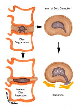 The process of disk degeneration following internal disk disruption and herniation.
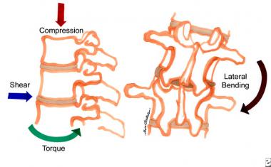 The various forces placed on the disks of the lumbar spine that can result in degenerative changes.
Of all connective tissues, the intervertebral disk undergoes the most serious age-related changes. By the third decade of life, the nucleus pulposus becomes replaced with fibrocartilage, and the distinction between the nucleus and the annulus becomes blurred. The proteoglycan, water, and noncollagenous protein concentrations decrease, while the collagen concentration increases. The increase in collagen concentration is more pronounced in the nucleus and in the posterior quadrants of the disk. It is more pronounced with age and moving caudally in the lumbar spine (similar to the Wolff law).
Biochemically, aging increases the ratio of keratin sulfate to chondroitin sulfate, and it also changes the proportion of chondroitin-4-sulfate to chondroitin-6-sulfate, with a parallel decrease in water content. Proteoglycan synthesis decreases, which decreases the osmotic swelling and the traffic of oxygen and nutrients to the disk. Because of this decreased traffic, breakdown products of link and noncollagenous proteins stagnate in the disk. Nonenzymatic glycosylation of these breakdown products accounts for the brown discoloration of the aging connective tissues.
Differentiating aging from degeneration is difficult. According to Pearce et al, "Aging and degeneration may represent successive stages within a single process that occurs in all individuals but at markedly different rates."[2] Aging and degeneration have in common decreased water and proteoglycan content in the disks, combined with increased collagen.
While sagittal alignment, facet joint arthritis, and genetics potentially play a role in intervertebral disk degeneration, the results of one study suggest that the rate of degeneration may be associated with age. Those of African ethnicity also showed a faster rate of degeneration when compared with whites; sex did not show a significant effect on degeneration.[3]
One study demonstrated that the presence of juvenile disk degeneration was strongly associated with overweight and obesity, low back pain, increased low back pain intensity, and diminished physical and social functioning. An elevated body mass index was significantly associated with increased severity of disk degeneration.[4]
Another study identified metabolic syndrome to be four times more prevalent in patients with radiographic evidence of severe degenerative disk disease as defined by degenerative spondylolisthesis or cervical or lumbar stenosis causing neurological symptoms[5] .
This cascade of degenerative changes can be subdivided into 3 stages: dysfunction, instability, and restabilization. The duration of the stages varies greatly, and distinguishing the signs and symptoms from one stage to the next is difficult.
Dysfunction involves outer annular tears and separation of the endplate, cartilage destruction, and facet synovial reaction. The symptoms of dysfunction are low back pain or neck pain, often localized but sometimes referred, and painful movement. The signs are local tenderness, contracted muscles, hypomobility, and painful extension of the back, neck, or both. Results of a neurological examination are usually normal.
The dysfunction stage is followed by the instability stage, in which disk resorption and loss of disk space height occur. Facet capsular laxity may develop, leading to subluxation. The symptoms are those of dysfunction (ie, "giving way" of the back, a "catch" in the back with movement, and pain with standing after flexion). The signs are abnormal movement (ie, during inspection or palpation), including observation of a catch, sway, or shift when standing erect after flexion. It has been found that the release of cytokines play a key role in all three stages and will likely be a target for therapeutic interventions in the future.
In the stage of restabilization, the progressive degenerative changes lead to osteophyte formation and stenosis. The symptoms are low back pain of decreasing severity. The signs are muscle tenderness, stiffness, reduced movement, and scoliosis.
NextLumbar surgery is indicated in patients with severe spinal stenosis, in those with intractable pain, and in patients in whom an appropriate 6- to 12-month nonoperative course of treatment fails. Surgery is elective, except in the presence of bowel and bladder symptoms or cauda equina syndrome.
In elective cases, other conservative modalities should have been tried and observed to fail. The patient should be prepared for the operation from a psychological viewpoint. Instability is present at 1 or 2 levels (as mentioned above in the instability phase of degenerative changes).
In the case of cervical disk disease with radiculopathy, the indications for surgical treatment are intractable pain, progressive motor or sensory deficit, or symptoms refractory in a reasonable period of nonoperative therapy. When the symptoms and signs correlate with radiographic evidence of root compression, various groups report a greater than 90% likelihood of a favorable outcome with both anterior and posterior approaches to surgery.
In the case of cervical disk disease with myelopathy, because the natural history is a stepwise worsening course, early surgery to decompress the spinal cord is recommended to arrest progression if the clinical and radiographic changes are well correlated. The best results for myelopathy occur when surgery is performed within 6 months of the onset of symptoms. Some series show improvement of greater than 70% with surgery in patients with myelopathy.
The spine is composed of 7 cervical, 12 thoracic, 5 lumbar, and a fused set of sacral and vestigial coccygeal vertebrae. Spine stability is the result of 3 columns in 1 as described by Dennis. Fracture or loss of 2 columns results in instability. The anterior column consists of the anterior longitudinal ligament and the anterior portion of the vertebral body. The middle column consists of the posterior wall of the vertebral body and the posterior longitudinal ligament. The posterior column is formed by the posterior bony arch; this consists of transverse processes, facets, laminae, and spinous processes.
Intervertebral disks form one quarter of the total length of the spinal column. Each vertebra has the potential for 6° of freedom, translation in all 3 axes of movement, and rotation around each axis. Not all vertebrae are created equal; the cervical vertebrae have the greatest freedom of flexion, extension, lateral rotation, and lateral flexion. This is because they are larger, they have concave lower and convex upper vertebral body surfaces, and they have transversely aligned facet joints.
Thoracic vertebrae have restricted flexion, extension, and rotation but freer lateral flexion because they are attached to the rib cage, are smaller, have flatter vertebral surfaces, have frontally aligned facet joints, and have larger overlapped spinous processes. The lumbar spine has good flexion and extension and free lateral flexion because its disks are large, the spinous processes are posteriorly directed, and the facet joints are sagittally directed. Lateral lumbar rotation is limited because of facet alignment.
The sensory of intervertebral discs is complex and varies depending on their location within the spinal column. In the cervical spine, studies by Bogduk[6] and Mendel[7] have demonstrated the presence of both nerve fibers as well as mechanoreceptors within the anulus fibrosus. Impulses from these structures are transmitted via the sinuvertebral nerves and branches of the vertebral nerves. In another study by Bogduk[8] , it was found that, like the cervical discs, the sensory innervation the lumbar intervertebral discs is derived from the sinuvertebral nerves but also from branches of the ventral primary rami and rami communicantes.
Relative contraindications to spinal fusion include smoking, morbid obesity, active infection, and severe medical or psychological problems.
Workup
Copyright © www.orthopaedics.win Bone Health All Rights Reserved