The term hemangiopericytoma (HPC) was first used by Stout and Murray in 1942 to describe a distinct neoplasm of pericytic origin.[1] However, over time, the staghorn-branching vascular pattern representative of HPC was found to be present at least focally in 15% of all soft-tissue tumors and was more of a characteristic histopathologic pattern than a specific clinicopathologic entity.[2]
Today, the diagnosis of HPC is primarily reserved to neuropathologists. The term solitary fibrous tumor (SFT) is favored by soft-tissue pathologists to describe a rare, heterogeneous group of benign and malignant neoplasms along a morphologic continuum.[3]
NextSFT was first described in 1870 by Wagner[4] and further established in 1931 by Klemperer and Rabin as a pleural neoplasm.[5] Three classical clinical forms of this entity are recognized, as follows[3] :
Soft-tissue SFT occurs across a histopathologic spectrum. On one end, a fibrous form is characterized by hyalinized, thick-walled vessels with opened lumina and strong CD34 reactivity. On the other end, a cellular form, representing the conventional HPC, has branching, thin-walled vessels and focal or absent CD34 reactivity.[2]
Lesions formerly known as HPC have been partitioned into three main groups. So-called true HPCs, with clear evidence of myoid and pericytic differentiation, include a subset of sinonasal HPC, subcutaneous infantile myofibromatosis or infantile HPC, and glomangiopericytoma or myopericytoma.[2] Many lesions have HPC-like features and are sometimes miscategorized. Most notably, the monophasic spindle cell or fibroblastic synovial sarcoma variant can be confused with HPC.[6]
The third group includes conventional SFT, a fat-forming SFT or lipomatous HPC, and a giant-cell–rich variant of SFT or giant cell angiofibroma.[2] This article focuses on the cellular variant of conventional SFT, or the previously termed conventional HPC.
Most cases occur in adults, with a median age of 45-50 years. SFT is less common in infants and children. Soft-tissue SFTs represent only about 1-2% of all soft-tissue tumors.[3]
The etiology is unknown.
SFTs are malignant tumors of mesenchymal origin that occur in the extremities. This tumor typically spreads via hematogenous dissemination, primarily to the lungs, but rarely spreads via the lymphatics. Metastatic disease is usually the cause of death.
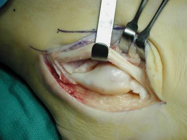
SFTs typically present as a deep, painless enlarging mass in the thigh, axilla or pelvis. Males and females are equally affected; the median age is 45 years.[3, 6] Patients may exhibit symptoms associated with a local compressive effect on viscera or neurovascular structures. Paraneoplastic syndromes such as hypoglycemia have been described.[7]
Workup
Copyright © www.orthopaedics.win Bone Health All Rights Reserved