The Lisfranc joint, which represents the articulation between the midfoot and forefoot, is composed of the five tarsometatarsal (TMT) joints. Jacques Lisfranc de Saint-Martin (1790-1847), a field surgeon in Napoleon's army serving on the Russian front, described a new amputation technique across the five TMT joints—one that did not require any bony osteotomy—as a swift solution to forefoot gangrene secondary to frostbite. This anatomic landmark became known as the Lisfranc joint, a term that is used today in the description of a wide spectrum of traumatic injuries to the TMT area of the foot.
The Lisfranc ligament is a solitary ligament that connects the first ray (first metatarsal-medial cuneiform articulation) to the middle and lateral columns of the foot. It is attached to the lateral margin of the medial cuneiform and the medial and plantar surface of the second metatarsal (MT) base. This is the only ligamentous support between the first ray and the second ray at midfoot level.
Although Lisfranc described the joint that bears his name, he did not actually describe the injury pattern well known by this eponym. As currently understood, a Lisfranc injury encompasses everything from a sprain to a complete disruption of normal anatomy through the TMT joints. This type of injury was later described in equestrian riders who got their foot caught in a stirrup when they fell from a horse.
Lisfranc joint injuries are rare, complex, and often misdiagnosed or inadequately treated. Injuries to the Lisfranc articulations frequently lead not only to arthritis but also to severe pain. Injury to the Lisfranc ligament, even in isolation, will result in functional instability with loss of longitudinal and transverse arch[1] ; early recognition and treatment of injuries to this ligament are important for preserving normal foot biomechanics and function. Lisfranc fracture dislocations and sprains carry a high risk of chronic secondary disability. Best outcomes for these injuries require prompt recognition and then anatomic reduction and stabilization.
NextThe Lisfranc joint is composed of five TMT joints in which the first through third MTs articulate with their corresponding medial, middle, and lateral cuneiforms, whereas the fourth and fifth MTs articulate with the cuboid. Functionally, the Lisfranc joint can be divided longitudinally into three columns, as follows:
A transverse line through these joints is not straight but highlights a recess, termed the keystone (much as in a Roman arch), that is formed by the second TMT joint. This joint lies approximately 1 cm proximal to the first TMT joint line and 0.5 cm proximal to the third TMT joint line.
The joints are bound by thick plantar ligaments that form an interlocking pattern between the tarsal and lesser MT bones 2-5. These are reinforced by attachments of the posterior tibialis tendon. The first TMT joint also has strong plantar ligaments across the joint; these are reinforced by the attachment of the peroneus longus and anterior tibialis tendons.
Also present between the lesser MTs is a series of intermetatarsal ligaments, which force the group to function more as a unit. No intermetatarsal ligaments exist between the first and second MTs, which is why they often exhibit divergent behavior. The weaker dorsal ligaments explain the majority of dorsal dislocations.[2]
The Lisfranc ligament originates from the plantar lateral aspect of the medial cuneiform and attaches to the plantar medial aspect of the second MT base. It is the thickest of the ligaments in this region, measuring up to 1 cm wide. This ligament provides the only soft-tissue link between the medial ray and the lesser MT and is responsible for this area's stability.
Motion at the TMT joints is variable. The second and third joints are the stiffest, with minimal motion in the dorsal or plantar plane and none in the medial or lateral plane. The third and first TMT joints exhibit progressively more motion in both planes but still are relatively stiff and mainly function as areas of adjustment to allow the MT heads to share weight equally.
The lateral two TMT joints demonstrate roughly three times more motion in the dorsal or plantar plane than the first TMT joint does. That motion is significant in the function of the foot and must be preserved to maintain normal function, especially if stiffness occurs in the medial and middle columns.
In the column theory, the middle column is more important for rigidity, and the medial and lateral columns are more important for shock absorption during gait. The lateral joints are more important for their mobile contributions to the balancing of forefoot weightbearing. This principle is important in treating these injuries.
In diabetic patients with neuropathy or those with idiopathic insensate feet, subacute diastasis can occur over time without notable pain. The absence of pain allows this gradual process to continue, so that a minor injury can lead to a Lisfranc injury.
In the authors’ opinion, the hallmark of an impending Lisfranc injury is the loss of the recess of the second MT base with the cuneiform, also known as the keystone. Radiographs are considered abnormal when weightbearing anteroposterior (AP) views of the foot show the first TMT joint to be at the same level as the second TMT joint, indicating proximal migration of the first ray (see the image below).
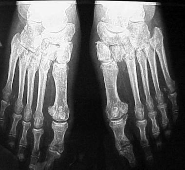 Radiograph illustrating diabetic patient with first ray instability of the right foot. The articular surfaces of the second and first metatarsal are level in the transverse plane, indicating proximal migration of the first ray. The left foot shows the advanced stage of an untreated Lisfranc injury with similar first ray instability.
The two major causes of Lisfranc injuries are as follows[3] :
In low-energy settings, TMT injuries are caused by a direct blow to the joint or by axial loading along the MT, either with medially or laterally directed rotational forces. In high-energy injuries, the method of loading is not significantly different, but the energy absorbed by the articulations results in significantly more collateral damage to bony and soft-tissue structures, creating such injuries as MT fractures, cuneiform instabilities, and cuboid fractures.
The damage to the tight ligamentous structures of this joint complex creates an unstable foot for weightbearing. The sense of instability and pain can occur whether or not overt evidence of instability is present. Chronic sprains resulting from relatively minor trauma can be the most debilitating sprains as a consequence of pain with weightbearing.
Lisfranc injuries account for 0.2% of all fractures.[4] The reported incidence of this uncommon injury is approximately 1 per 55,000 persons per year. It can occur in all ages but is more common in the third decade and is more common in males.[5] Subtle Lisfranc sprain and diastasis have become more commonly diagnosed in athletes.[6, 7]
Stable anatomic alignment is the best predictor of outcome.[8] The presence of fractures and/or articular destruction leads to poorer results, regardless of alignment. The incidence of posttraumatic arthritis reportedly ranges from zero to 58%.[9] One study reported that as many as 25% of patients develop posttraumatic arthritis even after fixation. This same study showed that there was no difference between acute and delayed (>6 weeks) surgical fixation. Purely ligamentous injuries seemed to have poorer outcomes.
Good results are achieved with open reduction and internal fixation (ORIF) at up to 6 weeks, but poor outcomes are seen after this time, arising from articular destruction, malalignment, and poor soft-tissue envelope.
Schepers et al performed a pedobarographic study that showed reduced contact time and reduced contact area of the forefoot in 26 patients relative to the uninvolved side.[10] Whereas these patients expressed good satisfaction with the procedure (primary ORIF [PORIF]), with Short Form (SF)-36 scores averaging 101, their functional scores were only fair, with a median American Orthopaedic Foot and Ankle Score (AOFAS) of 72 and a Visual Analog Scale (VAS) score of 7.
Clinical Presentation
Copyright © www.orthopaedics.win Bone Health All Rights Reserved