Scoliosis is a common deformity in many types of neuromuscular diseases (see the image below). It is generally most severe in nonambulatory patients. Severe curves of the vertebral column cause difficulties in sitting. Bracing neuromuscular curves does not affect the natural history of scoliosis and is not definitive treatment. Surgical stabilization constitutes the mainstay of treatment for neuromuscular scoliosis. Progressive curves require surgical correction and stabilization.
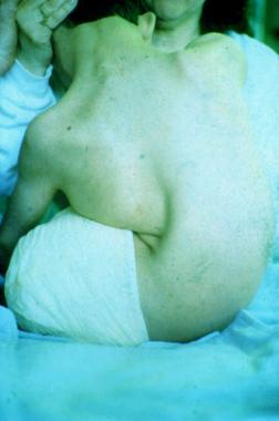 Neuromuscular scoliosis. Preoperative clinical picture of a young male with severe scoliosis secondary to quadriplegic cerebral palsy. Next
Neuromuscular scoliosis can be defined as a coronal and sagittal plane deformity of the spine in patients with abnormalities of the myoneural pathways of the body. In neuromuscular spinal deformities, progression occurs much more frequently than in idiopathic scoliosis.
In addition, progression often continues into adulthood. The long-term effects of the spinal deformity in patients with neuromuscular conditions can be disabling. Loss of the ability to sit occurs, as does an accompanying decrease in overall function. In addition, pulmonary function is markedly affected.
Because neuromuscular scoliosis has so many causes, the patterns and incidence vary greatly. However, the prevalence of spinal deformity in the patient with a neuromuscular disorder is much higher than in the general population. It ranges from 20% in children with cerebral palsy to 60% in patients with myelodysplasia. The prevalence rises to 90% in males with Duchenne muscular dystrophy. In general, the greater the neuromuscular involvement, the greater the likelihood and severity of scoliosis.
Scoliosis associated with neuromuscular disorders has been classified by the Scoliosis Research Society into neuropathic and myopathic types.
The neuropathic conditions have been subdivided into those with upper and lower motor neuron lesions. The group with upper motor neuron lesions includes diseases such as cerebral palsy, syringomyelia, and spinal cord trauma; the group with lower motor neuron lesions includes poliomyelitis and spinal muscular atrophy. The myopathic conditions include arthrogryposis, muscular dystrophy, and other forms of myopathy.
{refIntroductionPathophysiology}[[1] 1] The pathophysiology is not well understood. It seems logical to assume that scoliosis in these conditions is caused by muscle weakness, but this conclusion is difficult to support because some conditions are accompanied by spasticity and others by flaccidity. Furthermore, no consistent pattern of scoliosis is associated with a particular pattern of weakness.
The evaluation of a patient with neuromuscular scoliosis entails a thorough assessment of all body systems. Accurate diagnosis of the underlying disease entity is essential and may require muscle biopsy.
Assessing nutritional status and pulmonary function is extremely important. The child's caregivers should be interviewed to gain an appreciation of the patient's functional level. The orthopedic examination includes assessment of all extremities and joints for contractures.[2] Spinal deformity, decompensation, and shoulder balance are documented. Ambulatory status is also evaluated, and patients are classified as walkers, sitters, or nonsitters.
The two main indications for surgery are curve progression and deterioration in sitting ability.
The main contraindication would be inability to tolerate the procedure. A preoperative assessment of respiratory competency, cardiac status, nutrition, possible feeding difficulties, seizure disorders, urologic status, and metabolic bone disease is necessary to ensure that the patient is healthy enough to tolerate surgery.
Understanding the anatomy of the spine is crucial for safe and efficient exposure with a posterior approach. The incision is made from the spinous process above the most proximal vertebra to be instrumented to the most caudal extent of the proposed instrumented area. Identifying and staying in the midline is important so that muscle is not cut, which would lead to bleeding. The midline is identified by a thin line, which is actually the interspinous ligaments connecting the spinous processes.
Each vertebral level is exposed in a similar manner. An elevator is used to pull the soft tissue off of the spinous process, lamina, and transverse process of each respective level. To minimize blood loss, expose each segment completely the first time; do not leave soft tissue on the bone that will have to be removed later.
Workup
Copyright © www.orthopaedics.win Bone Health All Rights Reserved