The upper cervical spine is defined by the two most cephalad cervical vertebrae, C1 (the atlas) and C2 (the axis). This region is distinct in anatomic shape and is more mobile than the lower cervical spine, the subaxial cervical spine. The occipital condyles of the head (or the globe) rest upon the lateral masses of C1 (the atlas). These articular facets allow most of the flexion and extension of the head on the neck as the occipital condyles articulate on the atlas.[1, 2, 3, 4]
The ring of C1 has no vertebral body; the vertebral body that would correspond to C1 is connected or contiguous with the vertebral body of C2 and projects up as the dens (the tooth), also known as the odontoid of C2 (the axis). Most of the lateral rotation of the neck actually occurs at the C1-2 junction; the remaining motion of the cervical spine is distributed among the subaxial spine vertebral motion segments as a fractional amount (~7%) per level and is less in total than the C1-2 lateral rotation.
This area of the upper cervical spine is extremely mobile, and its stability is dependent on ligamentous structures. In unresponsive patients or those who are unable to report symptoms or pain, a C1 fracture or an occipital cervical dislocation must be excluded by radiographic screening. Also, displacement of the C1 ring may occur if the capsule or ligaments are disrupted, even without a C1 fracture; hence, the head may be displaced on the neck, and the atlas may also rotate around the odontoid or sustain a fracture of the dens.
NextJefferson originally described this type of C1 fracture in 1920.[5] The principal treatment is with a halo and vest or cast, which remains an effective current treatment for many of these fractures.
The C1 vertebra (atlas) is a closed ring. A fracture of a closed ring necessarily results in at least two areas of ring disruption. These disruptions are customarily accompanied by a spread of the C1 ring fragments as a result of the axial loading mechanism of this injury and the weight of the head.
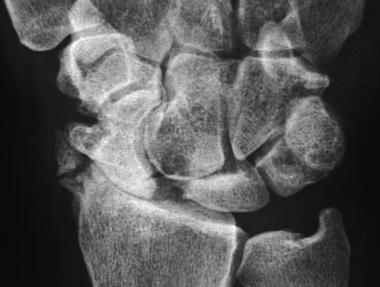
In addition to anteroposterior and lateral views, radiographs of the upper cervical spine include the open mouth view. This view may identify spreading or widening of the lateral masses or asymmetry of the separation of the odontoid from the lateral masses, which, in an appropriately centered radiograph, may be consistent with spreading of the C1 ring or a C1 fracture. Increased overhang of the lateral masses over the C2 facet totaling more than 6.9 mm suggests a fracture with disruption of the transverse odontoid ligament that may otherwise constrain displacement.
Fractures of the ring of C1 may be associated with an odontoid fracture; thus, the combination of the two fractures should be considered. Furthermore, congenital anomalies of the arch (eg, agenesis of the posterior ring) may be present. Anterior subluxation of C1 on C2 may be present and, if so, often indicates a disruption of the transverse odontoid ligament.
Fractures of the atlas account for 25% of atlantoaxial complex bony injuries, 10% of cervical spine injuries, and 2% of all spine injuries. Injury to the cervical spine occurs infrequently in pediatric populations, and although C1 represents only 1-2% of pediatric trauma and 2-10% of all cervical injuries in this population, the associated mortality is 16%.
The Jefferson fracture most commonly occurs as the result of axial loading on the head through the occiput, leading to a burst-type fracture of C1. Diving is the most frequent cause of this fracture, when it results from striking the head on an obstacle in shallow water; hence, the national program "Feet first, first time" (North American Spine Society, 2005) provides a motto for diving in unknown waters or shallow collections of water and has been an effective deterrent.
The next most frequent cause of this fracture is being thrown up against the roof of a motor vehicle, a car or bus, or even an aircraft, and the forces are distributed to the body through the neck. The third most frequent cause of these injuries is falls onto the head, except in toddlers, who are predisposed to injury from falls because of their disproportionate head size.[1]
Less frequently, when a significant rotatory force is exerted, an atlanto-occipital junction dislocation may occur, or the force may also be dissipated through the odontoid as an associated fracture.
The ring of C1 is a structural member of the cervical spine. Because it is a ring and because fracture results in disruption of this ring, more than one location is affected.
The fragments have a propensity to shift laterally, from both the weight of the head and the muscular contraction acting through this articulation; thus, occipital condylar support for the head is lost. The absence of the rigid bony structure and the lack of interconnection or interrelation of the attached ligamentous structures meet the definition of instability, particularly as the bony protective function of C1 for the neural elements is lost.
Vertebral artery injuries have been reported as a result of C1 fractures, especially with atlanto-occipital dislocations; small excursions of displacement can be fatal. In addition, vertebral artery injuries can occur and have been reported in the absence of severe trauma as a result of cervical traction, chiropractic manipulation, overhead work, or yoga exercises. Hyperextension is customarily accompanied with rotation; when this is not limited by normal restraints, it becomes excessive, severely diminishing blood flow through the vertebral arteries.
This diminished blood flow is particularly a problem in the posterior inferior cerebellar artery and may result in Wallenberg syndrome, which is characterized by ipsilateral loss of cranial nerves V, IX, X, and XI with cerebellar ataxia.
Horner syndrome may occur and, in some cases, may involve contralateral loss of pain and temperature sensation; involvement can extend up from a lateral medullary infarct and spread to the basilar superior cerebellar or the inferior cerebral artery, leading to sudden death, quadriplegia, and the locked-in syndrome, in which quadriplegia occurs with loss of lower cranial nerves and only eye-blinking is possible.
Patients customarily present with a history of trauma and a symptom of pain in the neck. Amid the massive number of patients who qualify as having this history and symptom, a few patients have an unstable C1 injury and may present neurologically intact, but they are at grave risk for neurologic compromise if not promptly diagnosed and appropriately stabilized and treated.
Patients with a complete spinal cord injury and no neurologic function continue to have only sensation on the face and motor control of the facial muscles from the cranial nerves. A tracheostomy is essential; the patient requires respiratory assistance and a volume respirator. If the C3-5 area is intact, the phrenic nerve may often be stimulated to contract the diaphragm. If stimulation of the phrenic nerve does not contract the diaphragm, then the spinal cord is no longer functioning; the cell body is dead, and a phrenic electrical stimulator is not effective.
Recognition and identification of a Jefferson fracture is the indication for treatment. Treatment consists of spinal stabilization to protect the patient from further nerve damage, including that to the brainstem. Children may represent less unstable cases, presumably because of periosteal stability, and they are often treated with a collar. The body of C1 is not visible radiographically until age 1 year.
Even in the absence of a C1 fracture, assessment of stability must include the associated structures. An atlanto-occipital dislocation or disruption and C1-2 instability, particularly when the transverse ligament may be disrupted, poses severe risk to the brainstem and upper spinal cord. Furthermore, with a C1 fracture, associations exist with unstable injuries such as odontoid fractures and other injuries to the upper cervical spine. In addition, the odontoid fragment may migrate into the foramen magnum, endangering the brainstem and upper spinal cord.
Specific treatment should be based on analysis of the mechanism and extent of the injury. In a younger patient with limited displacement of the C1, immobilization with a collar or halo and vest may be adequate.
In more severe cases, particularly with associated injuries such as odontoid fracture, bypassing the C1 ring with an occipital-to-cervical fusion extending to C2 or lower may be necessary. Instrumentation spanning that area may stabilize the C1 ring, which otherwise cannot easily be addressed directly, because both the anterior and posterior components of the ring are disconnected by the fracture and are not amenable to instrumentation or direct repair.
The care of any fracture requires attention to the joint above and below. This cervical complex has often been treated as two separable articulations, C0-1 and C1-2, but the three-unit occipitoatlantoaxial complex (C0-C1-C2) articulation is much more functionally relevant.
The significance is the proximity to the brain, brainstem, and upper cervical spinal cord, but that is contrasted with the very significant motion that occurs in this area. Although patients are routinely asked to flex and extend their necks to determine range of motion, some of that motion observed is between the occiput and the atlas, and as the patient rotates laterally, at least 50% of that motion is atlantoaxial.
The stability of the injury depends on the ligaments between the bony structures. On the frontal view, the projecting occipital condyles are supported by the lateral masses (observed as wedges, narrow medially and expanding laterally), resting on the corresponding superior articular surface of the axis (C2). Consequently, the lateral masses provide inherent stability because of this bony shape and also illustrate the extent of the instability when this bony structure is disrupted, particularly when these wedges displace laterally.
The projecting condyles of the occiput are stabilized with the occipitoatlantal capsule, as well as anterior and posterior atlanto-occipital membranes. The ligamentum nuchae is a significant stabilizing structure; its specific relevance to the atlanto-occipital axial complex is controversial but should be considered. Connections from the occiput to the axis are the tectorium membrane and the alar and apical ligaments, which do not appear to be bulky enough to be independently significant restraints.
The dentate ligaments (ie, the alar ligament and the apical ligaments) attach to the dorsal lateral surface of the dens and run obliquely to the medial surfaces of the occipital condyles. In 1974, Anderson and D'Alonzo classified a type 1 odontoid fracture as an avulsion fracture of the odontoid tip caused by the apical ligament, suggesting that these ligaments impart a significant degree of stability. A modified, treatment-oriented classification of odontoid fractures has been presented to expand the Anderson-D'Alonzo classification, but further assessment is needed.[6]
The transverse ligament goes from the medial surface of one side of the atlas to the other side and essentially constrains the axis to rotate around the odontoid in a closed ring of bone and the transverse ligament. As a consequence, the atlas can displace and embarrass the brainstem and spinal cord if this ligament ruptures or if an associated fracture of the odontoid is present as a result of this specific anatomic arrangement.
No significant contraindications to treatment exist, because the lack of stabilization, which commonly is initially provided either with traction or with a halo brace, can have fatal consequences. Any contraindications are mitigated by the potential for serious and even fatal neurologic consequences without treatment, as well as by the observation that halo with vest or traction can be relatively effective in immobilizing the upper cervical spine, with low associated morbidity.
Workup
Copyright © www.orthopaedics.win Bone Health All Rights Reserved