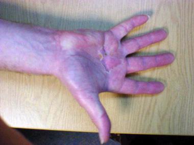
Since the advent of shoes, the fifth toe has been a source of discomfort for many people. Complaints typically involve poorly fitting shoes that create friction, irritation, and pain with each step. The problem can typically be solved conservatively with shoe modifications or proper foot maintenance; however, structural deformities of the toe often require surgical correction.
Fifth-toe deformities comprise several congenital and developmental problems that affect the fifth digit. Most are associated with contractures at the metatarsophalangeal joint (MTPJ) and the proximal interphalangeal joint (PIPJ), with or without varus rotation.[1]
Although fifth-toe deformities have long been recognized, correction of these deformities did not become prevalent until the early 20th century, when many authors began describing different aspects of the problem, along with surgical procedures to help correct them (see Treatment).
For patient education resources, see Corns and Calluses.
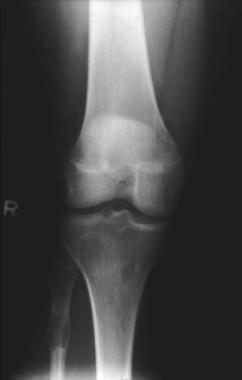
NextThree bones make up the fifth toe: the distal, middle, and proximal phalanges. They articulate together to make the distal interphalangeal joint (DIPJ) and the PIPJ. The proximal phalanx then articulates with the fifth metatarsal to make the fifth MTPJ. Medial and lateral condyles are present at the base of each phalanx, and epicondyles are present at the heads of the proximal and middle condyles.
A two-boned (biphalangeal) fifth toe has been reported in 37-76% of the population and involves a union of the distal and middle phalanges. When this occurs, the fifth toe is less flexible and often unable to accommodate pressure from standard shoes. This variant is more susceptible to irritation and may develop into a painful deformity.[2]
In a descriptive prospective study of 2494 feet in 1247 people, Gallart et al reported that a biphalangeal fifth toe was present in 46.3% of the feet and was bilateral in 97.4% of these cases.[3] The percentage of pathologic toes was significantly higher in patients with triphalangeal fifth toes (29.91%) than in those with biphalangeal toes (15.60%). The authors suggested that there may be an association between pathologic deviations and the greater mobility of triphalangeal fifth toes and that the greater rigidity of biphalangeal fifth toes may lead to lesser accommodation inside the shoe, which might result in less painful feet and decreased need for surgery.
The MTPJ has an extensor wing-and-sling mechanism that aids in extension of the digit. A slip of the extensor digitorum longus (EDL) to the fifth toe travels deep to the extensor wing and sling to insert into the dorsal aspect of the distal phalanx. No slip occurs from the extensor digitorum brevis (EDB) to the fifth toe; however, an occasional anomaly takes place in which an offshoot from the peroneus brevis tendon travels distal to insert into the dorsal-lateral aspect of the fifth MTPJ.
The fourth lumbrical muscle inserts into the plantar-medial fibers of the extensor wing to help adduct and plantarflex the proximal phalanx. The intrinsic third plantar interosseous and flexor digiti quinti brevis muscles insert into the plantar-medial and lateral aspects of the proximal phalanx respectively and function to stabilize the MTPJ against the stronger extrinsic flexor digitorum longus (FDL) and EDL. The abductor digiti minimi originates from the calcaneus and inserts into the plantar-lateral aspect of the proximal phalanx to place an abductory force on the toe.
The final two muscles to affect the fifth digit are the FDL and the flexor digitorum brevis (FDB), both of which plantarflex the toe. The FDL is deep to the FDB until the PIPJ, where the FDB splits, allowing the FDL to become superficial and continue distally to insert into the plantar portion of the distal phalanx. The FDB then rejoins to insert into the plantar aspect of the middle phalanx.
Clinical Presentation
Copyright © www.orthopaedics.win Bone Health All Rights Reserved