The occiput, the atlas (C1), and the axis (C2) form a functional unit, the occipitocervical articulation, which is characterized by a high degree of mobility and little intrinsic bony stability. Strong ligaments facilitate movement and keep these structures in place. Head rotation occurs primarily at this junction, and the odontoid bone is the axis that allows this rotation.[1, 2, 3, 4]
The atlanto-occipital joints allow movement in extension and flexion. In flexion, an anterior translation of C1 on C2, which normally does not exceed 3 mm in adults, exists. In children younger than 8 years, this translation can be as wide as 5 mm. In pathologic conditions (eg, abnormalities of the odontoid bone or in the ligaments that keep these joints together), this displacement increases and bone structures can pressure the spinal cord, producing clinical symptoms.
In the United States, atlantoaxial instability (AAI) with or without subluxation has been reported in as many as 10-30% of individuals with Down syndrome.[5, 6, 7]
In most instances, the radiologic findings are not associated with clinical symptoms.[8] The condition does not necessarily occur more frequently in the United States, although most cases reported to date have occurred in the United States.
No racial predilection is known for atlantoaxial instability in Down syndrome, and the role of sex is unclear. Most studies suggest a female preponderance[5, 6, 9] ; however, some reports noted a male preponderance,[10] and others found no difference between men and women.[11]
Most cases of atlantoaxial instability have been described in children.[12, 13] Longitudinal studies of children and adults show a high degree of stability both clinically and radiologically.[5, 8] In some individuals with radiologic evidence of atlantoaxial instability at the beginning of the study, radiographic findings normalized in subsequent evaluations.
A negative correlation exists between atlanto-odontoid distance and age.[14]
The following radiographs are provided for comparison between an image with a normal atlantoaxial relationship and that of a mild abnormality in a patient with Down syndrome.
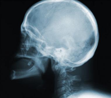
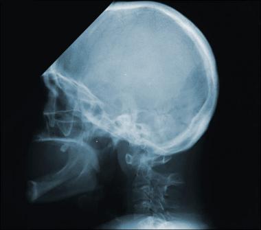
 This radiograph shows the normal relationships between the anterior arch of C1 and the odontoid bone, as well as the odontoid bone and the posterior arch of C1.
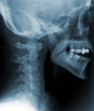
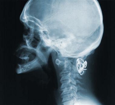
 Routine lateral cervical radiograph of a 28-year-old woman with Down syndrome confirmed by chromosomal analysis. The radiograph shows a mild degree of subluxation. The patient had no clinical signs of cord compression.
See also the following:
Many genetic and congenital developmental abnormalities affect the craniocervical junction,[1, 11, 15, 16, 17, 18] and a wide spectrum of congenital and acquired lesions can result in atlantoaxial instability (AAI).[19, 20]
In persons with Down syndrome, congenital absence or laxity of the transverse atlas ligament (which may be associated with congenital anomalies of the odontoid bone) must be considered. However, although this has not been proven to cause atlantoaxial instability[12, 9] and not every researcher agrees with this issue,[21] the excessive laxity of the posterior transverse ligament, which attaches the odontoid bone to C1, is considered an important factor in atlantoaxial instability.[12] When the transverse atlas ligament abnormality is present, trivial trauma to the cervical area or acute infections of the nasopharyngeal area may also produce subluxation.[22]
Other conditions are also known to present with generalized increased laxity of the ligaments (eg, Marfan syndrome, Ehlers-Danlos syndrome), and atlantoaxial instability might be a complication in these patients.[20] Additional factors may also be important; for example, malformations of the odontoid bone can be a factor in some cases.[22] Odontoid hypoplasia might be a predisposing factor by facilitating a slippage of the transverse ligament.
Lymphatic drainage of the craniocervical junction is the same as that of the cervico-occipital junction, and retrograde infections may play a role in the instability of the craniocervical joint.[22] The syndrome has been described after infections or surgical procedures (eg, mastoidectomies, tonsillectomies) in the mastoid, middle ear, and tonsils.[9]
Some pure cases of atlantoaxial instability are associated with degenerative arthritis or rheumatoid arthritis of the cervical spine.[23] Degenerative changes of the cervical spine are often overlooked, mostly because the majority of the studies of atlantoaxial instability are performed in children; however, these changes are frequently seen in adults with Down syndrome and might play a role in the development of clinical symptoms.[11]
Spinal canal stenosis due to C1 hypoplasia have been described in several children with Down syndrome and symptomatic atlantoaxial instability, which was minimal (only 6 mm) in some of them.[24] This paper reported that the cross-sectional area of C1 on computed tomography (CT) scan was significantly smaller in children with Down syndrome (505 mm2) when compared with children without trisomy 21 (602 mm2). This study showed that the space for the spinal cord can be reduced and a minimal degree of subluxation might be enough in some children to produce compression of the spinal cord.[24]
The early symptoms of subluxation are often subjective and difficult to elucidate in patients with mental retardation. This may result in a delay of diagnosis. In some cases, clinical symptoms must be inferred. For example, changes in behavior, refusal to participate in usual activities, changing hand preference, and urinary incontinence in previously continent individuals can be associated with atlantoaxial instability (AAI). No good longitudinal studies describe the natural evolution of asymptomatic persons with atlantoaxial instability; however, this condition is accepted as rarely symptomatic.
Suspect atlantoaxial instability in an individual with Down syndrome who presents with decreased motor skills or who develops torticollis, a gait disorder, or any form of progressive paralysis. Although the compression is in the high cervical spine, the first motor symptoms or signs may be discovered in the legs.
Signs and symptoms often result from mechanical compression of the cervical nerve roots and/or spinal cord.[25, 26] In some cases, the vertebral arteries may be distorted. Root compression at C1 and C2 levels produces pain in the upper cervical spine, the neck, and the occipital area that can extend to the head, eyes, ears, and/or throat.
Degenerative disease of the cervical spine, often seen in adults with Down syndrome, may complicate the clinical picture.[11, 13, 27, 28, 29]
Constant or intermittent vertebrobasilar insufficiency may produce dizziness, vertigo, tinnitus, diplopia, and/or syncope. Unilateral or bilateral tingling and numbness may be present.
In many instances, infections of the pharynx, the middle ear, and/or the upper respiratory tract precede symptoms of atlantoaxial instability. Clinical deterioration following any of these conditions justifies a complete evaluation.[22, 9, 30]
Motor system abnormalities are the most common findings at clinical presentation (primarily gait disorders and weakness).
Babinski sign and clonus in the lower extremities are not seen in healthy children with Down syndrome. If these signs are present, they should be considered abnormal. Progressive spasticity in the legs (characterized by increased muscle tone, muscle weakness, ataxic gait, increased deep tendon reflexes, Babinski sign, and clonus) can be a presenting sign.[26, 15, 31] In adults with Down syndrome who develop Alzheimer disease (also characterized by long-tract signs), these signs have less clinical value. (See Alzheimer Disease in Down Syndrome.)
In most instances, the neurologic signs and symptoms progress slowly in a matter of weeks. New-onset focal weakness might be an early sign of myelopathy due to atlantoaxial instability.[32]
Children with Down syndrome are hypotonic, and may remain hypotonic even with compression of the spinal cord.
Torticollis may be a presenting sign; the diagnosis of torticollis in a person with Down syndrome indicates atlantoaxial instability until proven otherwise.
Any pathology in the upper cervical spine can mimic symptoms of atlantoaxial instability (AAI). In addition, brainstem syndromes and cervical disk syndromes should be considered.
Adults with Down syndrome frequently develop Alzheimer disease. This disorder is associated with long-tract signs that may mask or mimic spinal cord compression.[5]
No specific laboratory tests are indicated in atlantoaxial instability (AAI). However, given the high frequency of hypothyroidism in individuals with Down syndrome, a full thyroid workup may be indicated. Hypothyroidism may play a role in this condition or be a separate cause of behavioral changes.[33]
Degenerative disease of the cervical spine is common in individuals with Down syndrome.[11] Subluxation may occur at multiple levels.[14]
Posterior atlanto-occipital subluxation with or without atlantoaxial instability has been described, including malformations of the odontoid bone, intervertebral disk abnormalities, and changes at the base of the skull.
Other conditions to consider include the following:
Neuroimaging studies of the cervical spine are the most important diagnostic tests.[8, 34] In most individuals with symptomatic atlantoaxial instability, the neurologic signs develop slowly, in a matter of weeks, so a neurologic examination before sport activities might be more predictive of atlantoaxial instability than radiologic screening.[9]
Lateral radiographs of the cervical spine in 3 positions (flexion, neutral, and extension) are used for screening purposes.[20] Even though new radiological techniques have been developed recently, the plain radiograph is still one of the best screening methods for early diagnosis and screening.[7] However, there are several unresolved issues with the use of cervical neck radiographs. How serious the risk is of asymptomatic instability is unclear. Serial radiographs may show the normalization of previous asymptomatic instability. In addition, there is no evidence that early screening may help to prevent symptomatic instability.[35]
A conservative approach consists of obtaining cervical radiographs in every child with Down syndrome, usually in early childhood and always before the child is involved in physical activities.[36] However, other authors do not agree with the need to perform regular radiologic screening evaluation.[21, 9]
Lateral radiographs of the neck are recommended by the Special Olympics Committee and were initially endorsed by the American Academy of Pediatrics (AAP)[37] ; however, the AAP later changed its position.[37] This resulted in heated arguments among experts in the field[6] and editorial reviews.[33] At the present time, this issue is still not fully resolved. National protocols have not been established and the recommendations regarding screening varies from state to state[17] and from country to country. For example, routine screening is not recommended in the United Kingdom.[38]
An unpublished longitudinal, clinical, and radiologic follow-up of 127 adults with Down syndrome in whom periodic cervical radiographs were obtained showed that the atlanto-odontoid junction remained stable with no significant radiologic changes in most of these individuals (N Alvarez, unpublished information, 2004). The individuals' average age at the time of the first set of radiographs was 38 years (range, 18-67 y) and an average age at the time of the last set of radiographs of 43 years (range, 23-67 y). In addition, when radiologic changes were observed, they were of no clinical significance. This study suggested that in the absence of clinical indications, repeat cervical radiographs are not needed in asymptomatic adults with Down syndrome. (N Alvarez, unpublished information, 2004).
The usefulness of the lateral cervical radiographs in asymptomatic children has not been fully resolved. Until more evidence-based information is available, the author recommends obtaining lateral cervical radiographs around the age of 4-5 years in asymptomatic children with Down syndrome. Those with radiologic indications of atlantoaxial instability should be closely observed by a clinician expert in the early subtle signs of this condition. In these individuals, the parents should be educated in the early recognition of early signs of complicated atlantoaxial instability.
Radiographic findings
In cooperative individuals, an open-mouth view of the upper cervical spine can assess the odontoid bone.
Normally, neck flexion shows minimal separation between the odontoid and the atlas (anterior atlanto-odontoid distance [AAOD]). This separation should be less than 3 mm (see the image below).[6] In children younger than 15 years, up to 4 mm can be considered normal.[20, 39, 40]
This radiograph shows the normal relationships between the anterior arch of C1 and the odontoid bone, as well as the odontoid bone and the posterior arch of C1.
An AAOD between 3-5 mm is borderline, and distances of more than 6 mm are considered suggestive of atlantoaxial instability (see the following images). Of asymptomatic children with Down syndrome, 17% have an AAOD greater than 5 mm.
Routine lateral cervical radiograph of a 28-year-old woman with Down syndrome confirmed by chromosomal analysis. The radiograph shows a mild degree of subluxation. The patient had no clinical signs of cord compression.
 This radiograph is of the same patient shown in the previous image, taken 8 years later when the patient, a woman with Down syndrome confirmed by chromosomal analysis, was aged 36 years. The routine lateral cervical radiograph taken at age 28 years showed a mild degree of subluxation, but the patient had no clinical signs of cord compression. Further clinical and radiologic follow-up showed no change either in the clinical picture or in the radiographs. The current radiograph, taken in a fairly neutral position because the patient did not allow for good flexion and extension positions, shows that the anterior subluxation, approximately 7-8 mm between the anterior arch of C1 and the odontoid bone, is still present. The distance between the posterior arch of C1 and the odontoid bone is approximately 12 mm, allowing enough space for the cervical spine.
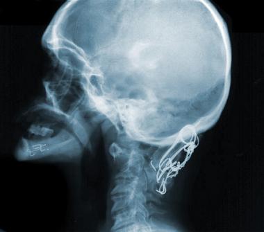
 Lateral cervical radiograph in a female with Down syndrome due to chromosomal translocation. The patient developed progressive gait deterioration, weakness, loss of muscle tone in the legs, and increased deep tendon reflexes in both arms and legs. Radiographs of the cervical spine documented a marked subluxation. She underwent fusion of the cervical spine.
 Lateral cervical radiograph in a female with Down syndrome due to chromosomal translocation (same patient as in previous image). The patient had developed progressive gait deterioration, weakness, loss of muscle tone in the legs, and increased deep tendon reflexes in both arms and legs. After undergoing fusion of the cervical spine, the follow-up radiograph revealed a distance between the anterior arch of C1 and the odontoid bone of almost 10 mm. The posterior distance was difficult to evaluate because of the wires. The radiograph seen here was taken after the patient underwent a second operation because of residual instability.
Measures of the posterior atlanto-odontoid distance (PAOD), which better correlate with the neural canal width, might have more clinical relevance than the measurements of the AAOD. Values of PAOD less than 12-13 mm are usually associated with clinical symptoms.
Computed tomography (CT) scanning and magnetic resonance imaging (MRI) are the best neuroradiologic tests to view the various structures of the cervical spine.[10] MRI is superior to CT scanning for evaluating the spinal cord, thus, MRI should be used when spinal cord compression is suspected. However, CT scans are useful as an adjunct to MRI for evaluation of bony abnormalities.
These imaging studies are indicated in all patients who show some clinical symptoms suggesting complications of atlantoaxial instability. They are optional in asymptomatic individuals when the cervical spine plain radiograph reveals subluxation greater than 5 mm. These studies are not indicated for routine screening.
Myelography is rarely indicated, as MRI resolution is sufficient in most clinical situations.
Medical (ie, nonsurgical) care for atlantoaxial instability (AAI) is primarily symptomatic to treat pain or discomfort. A cervical collar and traction can be tried, but if the patient's clinical symptoms do not improve, surgical treatment is indicated.[19, 20] A hard cervical collar might be needed after C1-C2 surgical fixation.[20] . In most instances, patients with Down syndrome and atlantoaxial instability do not need emergency surgery.
Regarding the indication of elective surgery, there is almost no disagreement that surgery is recommended in symptomatic patients and mostly when the signs and symptoms are suggestive of upper cervical cord compression. When patients are asymptomatic and the AAI is less than 4.5 mm, observation and continuation of regular activities is recommended. More active observation and limitation of activities like boxing, diving, and football, which may result in overflexion of the cervical spine, are indicated when the AAI is between 5.5 mm and 10 mm. Unless the patients are symptomatic clinical follow-ups are enough. When the AAI is 10 mm or more, surgery is commonly indicated. An algorithm has been developed to guide in the decision.[41]
The presurgical care is, in general, not different from the care needed for persons who do not have mental retardation.
Persons with Down syndrome have a higher incidence of congenital cardiac conditions, hepatitis, and hypothyroidism, and should be evaluated for these conditions before surgery. Emotional and behavioral issues could be complicating factors, mostly in the postoperative period. Explaining the inconveniences associated with major surgery to people with developmental disabilities is always difficult.
The anesthetist’s management of a person with atlantoaxial instability requires great care to avoid unnecessary mobilization of the neck that can occur to have access to an airway during the operation.[42, 43]
Consider consultation with a neurologist when the symptoms suggest atlantoaxial instability and diagnostic workup is needed. Neurosurgery or orthopedic specialists should be consulted when surgery is being considered.
Red flags (warning signs/symptoms) include the following:
In individuals with radiologic evidence of atlantoaxial instability without clinical signs or symptoms, activities that might produce injury to the cervical spine should be restricted.
Without evidence of subluxation, physical activity restriction is unnecessary.
Symptomatic individuals need full restriction of physical activities until surgery is performed.
Explain the following to parents and/or legal guardians:
The preferred surgical option for atlantoaxial instability (AAI), in the presence of symptoms, is fusion of C1 to C2.[20, 44] Surgery is more effective in individuals in whom atlantoaxial instability has been diagnosed recently. In those with long-standing symptoms, surgery may prevent further deterioration, but chronic symptoms do not improve significantly.[6]
Fusion of the cervical spine is not complication free. In some cases, the clinical situation deteriorates after surgery.[20] The C1-C2 fusion might lead to instability of the atlanto-occipital joint.[45]
When fusion has failed, a second corrective fusion is always an option. Alternative procedures that have been explored with some success are transoral odontoidectomy, endoscopic endonasal odontoidectomy,[46] and more recently, a single stage posterior approach.[47]
Persons with Down syndrome present a special problem when in need of operations that require tracheal intubation during anesthesia. The extension of the neck can result in compression of the spinal cord. Somatosensory and motor evoked potentials, usually not indicated in surgery that does not involve the spinal cord such as abdominal or thoracic or neck surgery, is an appropriate indication in these cases to protect complications of craniocervical instability.[48]
Pain should be expected in the postoperative period. In most instances, because of their mental retardation, patients with Down syndrome are not able to verbalize their pain, and behavioral changes might be the only indication of pain.
Companionship at the bedside is important for these patients. The physician should arrange for this companionship. The ideal situation is to have someone at the bedside 24 hours daily, preferably persons who know the patient well.
In most instances, a cervical collar is used to stabilize the surgical area. Because most of the patients have some degree of mental retardation, implementation of a cervical collar might require some sedation.
The main complication following surgery for atlantoaxial instability is spinal cord compression. In addition, root compression in the cervical area might be associated with posterior neck and occipital pain.
Torticollis may be a sign of subluxation.
Patients might need to be in bed for many days after the surgery, and deep vein thrombosis (DVT) may be a complication during this period. The caretakers should be instructed in prevention of DVT, such as moving the patient in bed and performing passive exercises of the legs. The rehabilitation should be started as soon as possible. Patients should be encouraged to move, even when in bed.
Other complications of the surgery, such as postoperative bleeding in the cervical spine, might result in compression of the upper cervical cord. This could lead to motor paralysis that might be very difficult to diagnose in the early stages. In extreme cases, respiratory depression might be a complication.
Manipulation of the cervical spine is always dangerous in individuals with Down syndrome (even those who are asymptomatic) with atlantoaxial instability (AAI).
Relaxation during anesthesia might allow extreme movements of the cervical spine. This should be a concern in any person with Down syndrome who needs general anesthesia; however, children with normal radiographic studies of the cervical spine can tolerate flexion and extension of the cervical spine during anesthesia.[49]
Before elective intubations, a cervical spine radiograph series should be performed. Fiberoptic intubation may be desirable in patients with Down syndrome to avoid overmanipulation of the neck.
Emergency intubation of people with Down syndrome should be performed with extreme care and minimal manipulation of the neck, mostly if the condition of the atlantoaxial instability is unknown.
Physical activities that result in trauma to the cervical spine should be avoided.
Following surgery, patients are likely to remain in the hospital for at least 1 week. Because sending these patients home is difficult, they might require transfer to a rehabilitation hospital. The average stay at the rehabilitation hospital is 1 month.
Nursing homes should be avoided, as they are usually not equipped for patients with Down syndrome.
Individuals with radiologic evidence of atlantoaxial instability but no symptoms should have at least a yearly clinical evaluation. Immediate reevaluation is needed if they present with new symptoms.
In children who have atlanto-odontoid distance (AAOD) of 3 mm or less, repeating the cervical radiograph is probably unnecessary. These children need clinical follow-up only. Neuroimaging should be pursued only if new neurologic signs arise.
No protocol exists for children with AAOD of 3-5 mm. Clinical follow-up and restricting activities that may traumatize the cervical spine (eg, somersaults, contact sports, trampoline exercises) may be all that is needed. Consider repeating the cervical radiograph in 1-2 years.
In patients in whom the AAOD is greater than 5 mm, a more complete evaluation with a cervical magnetic resonance imaging (MRI) is recommended. This also applies for children whose posterior atlanto-odontoid distance (PAOD) is less than 12 mm.
Only a small percentage of children with radiologic evidence of AAI will become symptomatic. The symptoms are often insidious. Quadriplegia is reported occasionally (but rarely) as an initial symptom.
Clinical deterioration can be observed after minimal trauma or after acute pharyngeal infections.
Atlantoaxial instability (AAI) does not usually produce clinical symptoms. In symptomatic individuals, treatment in the acute phase may reverse the symptoms. In long-standing cases, the surgical procedure is not associated with clinical improvement.
Several studies have shown that serious complications are rare.[33] The primary complications result from spinal cord compression. In most cases, signs and symptoms progress slowly. The diagnosis can be made, therefore, before the advanced stages of the disease.
Death is unusual but may occur in cases of acute decompensation as a result of respiratory arrest related to compression of the high cervical spinal cord.[50]
Copyright © www.orthopaedics.win Bone Health All Rights Reserved