Synostosis, or osseous union, of any two adjacent bones can involve any part of the upper extremity. In 1793, Sandifort provided the initial description of congenital radioulnar (radial-ulnar) synostosis. This condition is caused by a failure of segmentation between the radius and the ulna.[1, 2] Synostosis between the radius and ulna can take two general forms: congenital and posttraumatic. Each form may be further classified into types (see Presentation, Classification).
Posttraumatic radioulnar synostosis is a separate entity from the congenital form, having a different cause, treatment, and prognosis.[3] The traumatic form can occur anywhere between the radius and ulna along the length of the interosseous membrane. Gros first described posttraumatic radioulnar synostosis in 1864, reporting on a vicious union found in autopsy specimens. Groves later postulated that the success of treatment depended on where in the forearm synostosis had occurred.[4, 5]
NextThe skeletal anomaly includes varying degrees of proximal radial and ulnar fusion, with or without involvement of the radial head. If the radial head is involved, it may be dislocated anteriorly or posteriorly.[6] A fibrous synostosis may allow limited motion. Regional soft-tissue hypoplasia is often present in severe cases, including those in which atrophy and fibrosis of the brachioradialis, pronator teres, pronator quadratus, and supinator muscles occur. The interosseous membrane also may be abnormal.
Embryologically, the upper limb bud arises from the unsegmented body wall at 25-28 days. The elbow becomes visible at 34 days, and the humerus, radius, and ulna become visible at 37 days. Initially, the three cartilaginous analogues of the humerus, radius, and ulna are connected before segmentation. Therefore, for a short time, the radius and ulna share a common perichondrium. Abnormal events at this time can lead to a failure of segmentation. The duration and severity of the insult can determine the degree of subsequent synostosis.
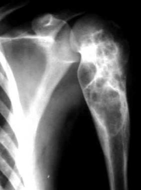
Endochondral ossification then proceeds, and the cartilaginous synostosis ossifies, either partially or completely, in the longitudinal or transverse plane. In the forearm, congenital radioulnar synostosis usually occurs between the proximal radius and the ulna. Although the condition is present at birth, it usually is not discovered until early adolescence, when the patient presents with a lack of pronation and supination.
Initially, the union may be more of a synchondrosis, but as the skeleton matures, the osseous bridge between the radius and ulna becomes more radiographically apparent. Usually, motion between the two adjacent bones, if existent, is minimal.[7, 8, 9, 10, 11, 12]
The most common cause of posttraumatic radioulnar synostosis is an operatively treated forearm fracture. Patients with high-energy comminuted open fractures appear to be more likely to develop this complication. Monteggia and proximal forearm fractures also appear to have a higher incidence of synostosis.[13] The use of bone graft and of screws protruding through the opposite cortex also increase the incidence of synostosis.
Additionally, radioulnar synostosis is described as a consequence of soft-tissue injury, reconstructive procedures, any trauma causing hematoma formation between the radius and ulna, or injury to the interosseous membrane.[14] Patients with closed head injuries (skull/cranial trauma) appear to be more prone to this complication, presumably for the same reason that they develop heterotopic ossification.[15, 16]
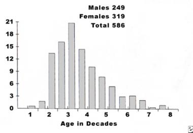
Congenital radioulnar synostosis occurs rarely, with approximately 350 cases reported in the literature. The rarity of this condition often leads to a delayed clinical diagnosis. Cleary and Omer reported an average patient age at diagnosis of 6 years, with a range of from 6 months to 22 years.[17] There is no sex predilection in congenital radioulnar synostosis, and no particular inheritance pattern is apparent. Sixty percent of cases are bilateral.
Because congenital radioulnar synostosis is caused by an in-utero insult, its association with other abnormalities is not surprising. About one third of cases are associated with general skeletal abnormalities, such as hip dislocation, knee anomalies, clubfoot, polydactyly, syndactyly, Madelung deformity, ligamentous laxity, thumb hypoplasia, carpal coalition, and problems of the cardiac, renal, neurologic, and GI systems.
Some associated abnormalities and syndromes are genetically determined, including acrocephalosyndactyly, Apert syndrome, Carpenter syndrome, arthrogryposis, mandibulofacial dysostosis, William syndrome, Klinefelter syndrome, Holt-Oram syndrome, microcephaly, multiple exostoses, and fetal alcohol syndrome.[18, 19] In 20% of their patients, Cleary and Omer found a genetic basis for an autosomal dominant form (with variable penetrance) of congenital radioulnar synostosis.[17]
Overall, results of surgical treatment for posttraumatic radioulnar synostosis are fair at best, with high failure rates and, commonly, loss of approximately one half of the intraoperative rotation. The use of postoperative indomethacin or low-dose limited field irradiation within the first 5 days after surgery has been shown to be effective in limiting the recurrence of synostosis.
Clinical Presentation
Copyright © www.orthopaedics.win Bone Health All Rights Reserved