Mannerfelt syndrome refers to rupture of the flexor pollicis longus tendon from attrition caused by a bony spur in the carpal tunnel. Such ruptures were described by multiple authors from 1891 on.[1, 2, 3, 4, 5, 6] In 1969, Mannerfelt published his series of attrition flexor tendon ruptures in rheumatoid arthritis (RA) caused by bony spurs in the carpal tunnel.[7] The flexor pollicis longus tendon was the flexor tendon most commonly ruptured in this setting; consequently, this specific lesion was named after Mannerfelt.
Functional loss in Mannerfelt syndrome is variable. Even if the tendon has ruptured, continuity may exist through the peritendinous sheath or a pseudotendon. The patient presents with weak or no flexion of the distal phalanx. Flexor tendon ruptures occur consecutively, starting with the flexor pollicis longus and thereafter affecting other flexor tendons in a more ulnar direction.[8] Bilateral ruptures have been reported.[9]
In his series of patients with affected flexor tendons in the carpal tunnel, Mannerfelt reported that 20 of 25 patients had involvement of the flexor pollicis longus tendon and that nearly all flexor tendon attritions occurred in women.[7]
NextThe flexor pollicis longus originates from the middle of the anterior surface of the radial shaft, the adjoining part of the interosseous membrane, and the coronoid process. It is the most radial tendon in the carpal tunnel. The tendon passes under the transverse carpal ligament and around the hook of the distal scaphoid, and it inserts into the base of the distal phalanx of the thumb, flexing the distal phalanx.
The anterior interosseous branch of the median nerve supplies the flexor pollicis longus. The muscle receives its blood supply from radial artery muscle perforators. Within the tendon sheath of the thumb are 2 distinct vincula. Feeding vessels to the trapezium and scaphoid are present on the volar radial aspect of the trapezioscaphoid joint. These vessels course along the trapezioscaphoid ligaments to reach the scaphoid tuberosity.
In rheumatoid arthritis (RA), according to Mannerfelt, the intercarpal ligaments lose strength, and the carpal flexors exert a continuous pull, causing volar subluxation of the carpal bones. Destructive rheumatoid inflammatory tissue follows the feeding vessels, causing erosions at the distal volar scaphoid and proximal volar trapezium.[10] Bony tissue between the erosions forms sharp spurs that cut through the weakened carpal tunnel floor and can lead to attrition of flexor tendons (see the image below).
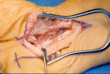 Mannerfelt syndrome. Edges of ruptured flexor pollicis longus tendon can be seen. Bony spur is present at floor of carpal tunnel.
The flexor pollicis longus tendon is usually ruptured by a scaphoid spur.[11] The site of the rupture is usually the scaphoid, followed, in descending order of frequency, by the trapezium, distal ulna, hamate, lunate, distal radius, and ulnar sesamoid.[12, 13, 14, 15, 16, 17, 18, 19]
The patient usually presents with a history of rheumatoid arthritis and loss of thumb interphalangeal joint flexion.
Because the flexor pollicis longus is the only muscle to flex the interphalangeal joint of the thumb, the Mannerfelt lesion is usually clinically apparent on physical examination. The examiner stabilizes the metacarpophalangeal joint, and the patient is asked to flex the interphalangeal joint. In the case of rupture, the patient is unable to flex the distal thumb phalanx or, at most, is able to flex it weakly (see the image below).
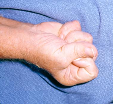 Mannerfelt syndrome. Patient is unable to flex interphalangeal joint of thumb after spontaneous rupture of flexor pollicis longus tendon.
Incomplete anterior interosseous nerve paralysis and rupture of the flexor pollicis longus tendon resemble each other clinically. A simple and reliable test can differentiate the 2 conditions.[20] The physician dorsiflexes the patient’s wrist. The thumb is hyperextended at both the carpometacarpal and metacarpophalangeal joints. If the flexor pollicis longus is intact but paralyzed, the interphalangeal joint flexes spontaneously and resists passive extension when tested. If the tendon is ruptured, the interphalangeal joint remains extended.[21]
Plain radiographs can reveal only gross radiocarpal changes. Because of wrist stiffness, an axial projection of the carpal tunnel is not easy to obtain.
Linear tomography does not show sufficient detail. Mannerfelt used circular tomography with an angle of 42°. The tomographic plane was perpendicular to the long axis of the forearm, and an axial projection of the carpal tunnel was obtained. This method revealed the carpal tunnel floor and the opposing aspects of the hamate hook and trapezium.
Magnetic resonance imaging (MRI) can accurately depict the level of rupture and the gap between the tendon ends. It also reveals the appearance of the adjacent tendons. Thus, MRI may assist the surgeon in choosing a treatment approach (eg, suture, graft, or tendon transfer).[22]
Medical management of an inflamed tenosynovium may reduce destructive pannus and spur formation and thereby also decrease the risk of tendon rupture. Once a Mannerfelt lesion is identified, surgical treatment is always recommended.
Before exploration, written informed consent should be obtained. The patient should be informed of the possibility that one of the reconstruction options described below will be needed. The patient should also be informed that the procedure does not cure the underlying problem (eg, rheumatoid arthritis [RA]). Therefore, recurrence of tenosynovitis and additional ruptures are possible.
Once a Mannerfelt lesion is identified, division of the transverse carpal ligament in rheumatoid hands is recommended. The surgeon should examine the carpal tunnel floor for bony spurs and excise them. In addition, full flexor tenosynovectomy should be performed. Early tenosynovectomy and removal of bone spurs can prevent tendon attrition.
Exploration of the carpal tunnel is recommended for tendon repair and prevention of further ruptures. Even in patients with a fused interphalangeal joint, the carpal tunnel should be explored because the bony spur that caused attrition of the flexor pollicis longus tendon may affect the remaining flexor tendons.
For a functional interphalangeal joint, tendon repair or reconstruction is recommended. When the distal tendon end is beyond the wrist and cannot be retrieved in the wound, a full-length tendon graft or tendon transfer is recommended. A nonfunctioning flexor pollicis longus muscle is an indication for tendon transfer. If the interphalangeal joint of the thumb is arthritic or unstable, fusion may be a better procedure for a functional thumb.
A linear incision is made over the 4th ray in the proximal palm and extended across the wrist in a zigzag manner, with care taken to avoid the motor branch and the palmar cutaneous branches of the median nerve. The bony spur is excised from the scaphoid or the trapezium. Adjacent soft tissues are mobilized to cover the exposed bone. The defect can be covered with a silicone membrane, a transverse carpal ligament flap, or a tendon graft. Repair can be done with a short (bridge) tendon graft, a standard full-length tendon graft, or a tendon transfer.
A short tendon graft is recommended when both tendon ends are at the wrist level. This short graft can be obtained from the palmaris longus, the flexor carpi radialis, or the abductor pollicis longus.[23, 24] When the distal tendon end is beyond the wrist and cannot be retrieved in the wound, a full-length tendon graft or tendon transfer is recommended.
For a full-length tendon graft, a volar zigzag incision is made on the thumb and extended proximally into the palm. The ruptured distal tendon part is resected, with care taken not to injure any of the pulleys. Through another incision at the distal forearm, the musculotendinous junction of the flexor pollicis longus is identified. The palmaris longus or plantaris tendon is harvested.
With a pediatric feeding tube or a tendon passer, the tendon graft is brought through the sheath and pulleys. The distal end of the graft is attached to the terminal phalanx with a pullout suture or suture anchor. Setting the tendon tension requires positioning the wrist in a neutral position, with the thumb opposed to the index finger and the thumb interphalangeal joint at 30° flexion. The proximal end of the graft is then woven into the flexor pollicis longus tendon.
After wound closure, the wrist is kept at 15-20° flexion and the thumb at 30° palmar abduction. A dorsal splint is applied, allowing passive flexion of both thumb joints. After 6 weeks, the pullout suture is removed.
A clear indication for a tendon transfer is a nonfunctioning flexor pollicis longus muscle. Compared with a tendon graft, a tendon transfer has fewer juncture sites (ie, only 1), yields a more viable tendon, requires no extra procedure for tendon graft harvest, and provides a motor in cases of direct flexor pollicis longus muscle damage or nerve injury. Possible disadvantages are proximal interphalangeal joint hyperextension and loss of flexor power in the donor finger.[25]
The flexor digitorum sublimis tendon of the long finger is used for a tendon transfer because it is longer than the rest of the flexor tendons and because the ring and small fingers are not impaired. The tendon is distally detached from the middle phalanx of the donor finger, passed under the median nerve, and brought through the tendon sheath and pulleys of the thumb by using a pediatric feeding tube or a tendon passer. The flexor pollicis longus stump is resected, and the underlying volar proximal surface of the distal phalanx is trimmed.
The flexor digitorum sublimis tendon is secured at the volar aspect of the distal phalanx of the thumb with a pullout suture. A bone anchor can be used instead, also placed in the volar proximal edge of the distal phalanx. A ring or small finger sublimis tendon transfer may be considered as an alternative. A flexor tendon tenosynovectomy is performed to prevent further ruptures. The wrist and thumb are immobilized in flexion for 3 weeks, after which passive and gentle active motion is allowed. If a pullout wire is used, it is removed after 6 weeks.
As noted, if the interphalangeal joint of the thumb is arthritic or unstable, fusion may be a better procedure for a functional thumb. Arthrodesis of the interphalangeal joint of the thumb can provide a strong pinch and improve function. Some authors recommend fusion at 0-5° flexion, whereas others prefer 15-20° flexion. Many fusion techniques with high success rates have been described.
Schneider and Wiltshire treated 14 patients who underwent ring-finger flexor digitorum sublimis transfer for the treatment of irreparable lesions of the flexor pollicis longus. Results, as measured by return of interphalangeal joint motion, were good in 12 patients and fair in 1 patient; failure occurred in 1 patient.[26]
Complications are minimal, though rupture of the tendon reconstruction or nonunion of an interphalangeal fusion can occur. Tenolysis may also be required.[27]
Copyright © www.orthopaedics.win Bone Health All Rights Reserved