Thumb function constitutes about 50% of hand function as a whole. The thumb metacarpal base is a unique joint that allows a wide range of motion while maintaining stability for grasp and pinch in a variety of positions.
The images below depict Rolando fractures.
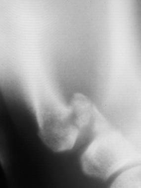 Lateral tomograph of a Rolando fracture clearly shows the varus angulation at the fracture, as well as the multiple fragments of the articular surface.
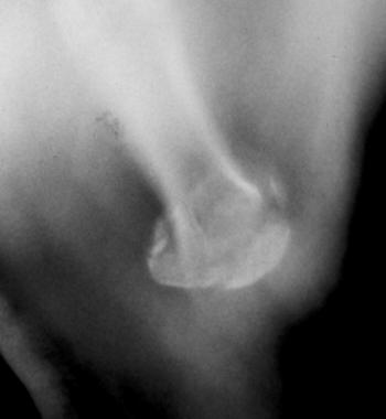 Anteroposterior tomograph of a Rolando fracture further emphasizes the extent of comminution of the articular surface (same patient as in Image above).
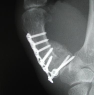 Radiograph of a healed Rolando fracture following fixation of the articular surface and neutralization with a small plate.
Multiple fracture patterns of the thumb base have been described, including juxta-articular metaphyseal fracture, Bennett fracture, and Rolando fracture. Interest in the fixation of these fractures has been stimulated by the marked decrease in hand function that can develop in the affected patients if disabling arthritis occurs in the thumb carpometacarpal articulation as a result of articular incongruity following such fractures.[1, 2, 3, 4, 5]
Metacarpal Fractures - Orthopedic Surgery
Thumb Fractures and Dislocations - Plastic Surgery
Thumb Dislocation Joint Reduction - Clinical Procedures
Metacarpal Fractures and Dislocations - Plastic Surgery
Bennett Fracture - Orthopedic Surgery
NextRolando fracture initially was described in 1910 in a series of 12 metacarpal base fractures, of which 3 involved a Y-shaped split of the joint surface.[6, 7] The fracture was described as having 3 major fragments: metacarpal shaft, dorsal metacarpal base, and volar metacarpal base. Currently, the term has come to include essentially all comminuted thumb metacarpal base fractures.[8]
The initial treatment options that were described mainly focused on closed treatment, either cast immobilization or a short period of splinting followed by early motion to mold joint surfaces. With the advent of internal fixation techniques, especially smaller implants, interest in operative treatment has increased over the past few decades.
Rolando fracture is analogous to the pilon fracture of the distal tibia and appears to be secondary to a significant axial load that splits and crushes the metacarpal articular surface. Rolando described 2 cases that occurred secondary to a fall on the radial side of the hand, with the thumb in adduction, and a third case that was caused by a closed fist, with the thumb folded and held in the palm, striking an adversary's head.[6]
Following an injury similar to that described above, the fracture is at risk of further displacement due to the resting tone present in the multiple tendons that act on the thumb. The extensor pollicis brevis and longus shorten the thumb ray, as does the pull of the flexor pollicis longus. The adductor pollicis muscle tends to pull the distal metacarpal toward the palm, which, in conjunction with the abductor pollicis longus acting on the metacarpal base, commonly produces varus at the metaphyseal-diaphyseal junction.
Following injury, patients present with a swollen, tender thumb base. If significant varus has developed, a clinically visible deformity may be present. However, swelling can mask a surprising amount of angulation. Neurovascular and tendon injuries are not commonly associated with this fracture.
Significant joint incongruity (ie, >1-2 mm of articular step-off) mandates treatment. However, the type of treatment can vary and is somewhat controversial. Large articular fragments in which screws can be used are probably best supported by plate and screw fixation, whereas massive comminution is best treated with a form of traction (see Future and Controversies). Open fractures require debridement, and operative stabilization is recommended to stabilize the skeleton and allow soft-tissue healing. Pin fixation or external fixation is preferred in the presence of open injuries to minimize soft-tissue stripping.[9]
The carpometacarpal joint surface consists of 2 reciprocal interlocked saddles that allow motion parallel and perpendicular to the plane of the palm. Compressive forces across the joint appear to be magnified during pinch and have been estimated at 12 times the pinch force.[10] Articular incongruity, therefore, is subjected to high forces and increases the likelihood of arthrosis development. As a result, interest has been spurred to improve the accuracy and security of reduction techniques.
Contraindications to surgery are few; a systemically ill patient following polytrauma who cannot undergo any surgical procedure is an example of a patient in whom surgery would be contraindicated. An open fracture that has large fragments (normally treated with plate and screw fixation) and is massively contaminated would best be managed with traction and repeated debridements.
Workup
Copyright © www.orthopaedics.win Bone Health All Rights Reserved