What Is Minimally Invasive Spine Surgery?
In essence, minimally invasive spine surgery is the performance of surgery through small incision(s), usually with the aid of endoscopic visualization (i.e., very small devices or cameras designed for viewing internal portions of the body).
Why Is Minimally Invasive Spine Surgery Needed?
Minimally invasive spine surgery has developed out of the desire to effectively treat disorders of the spinal discs with minimal muscle related injury, and with rapid recovery.
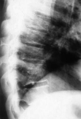
Traditionally, surgical approaches to the spine have necessitated prolonged recovery time. For example, in the 1990s the state-of-the-art procedure for fusion of the lumbosacral spine has been the instrumented posterolateral fusion. In order to perform this procedure, the back muscles are moved away from their spinal attachments, allowing the surgeon space to place rods, screws, and bone graft.
First, this surgical approach (i.e., dissecting the muscles) produces the majority of the perioperative pain and delays return to full activity. The degree of the perioperative pain necessitates the use of significant pain medication with their inherent side effects. Also, the degree of the perioperative pain delays return to normal daily activities and nonphysical work.
Second, the dissection of the paraspinal muscles from their normal anatomic points of attachment results in a healing by scarring of these muscles. The various layers of the individual muscle scar to one another losing their independent function.
In addition, it has been found that this type of dissection results in the loss of innervation (i.e., the supply of nerve stimulation) of the muscles with subsequent wasting away. A permanent weakness of the back muscles results. This weakness itself may be symptomatic (as a back fatigue-type pain) and/or limit the patient's function - particularly in those who perform physical work. These side effects of the posterior approach to the lumbar spine have been called fusion disease.
Clearly, with such significant muscle injury associated with surgical approaches to the spine, the need existed for the development of less invasive surgical techniques. It was envisioned that minimally invasive techniques would offer several advantages including: -Reduced surgical complications - Reduced surgical blood loss - Reduced use of postop narcotic pain medicines - Avoidance of fusion disease - Reduced length of hospital stay - Increased speed of functional return to daily activities The Emergence of Minimally Invasive Techniques With the advent of laparoscopic general surgery in the 1980s, other surgical specialties began searching for applications of the visualization technology. It became apparent that sections of the spine, such as the thoracic (chest) and lumbar (lower back) regions could be exposed using minimally invasive technology.
Development of Laparoscopic Approaches to the Lumbar Spine
During the 1980s, laparoscopic technology was developed that enabled exposure of the lumbar spine. Although visualization was possible, initially there was not a method of fixation of the lumbar motion segment which could be introduced via laparoscopic tubes, and that could provide stability comparable to posterior fixation. Without the ability to instrument the spine laparoscopically, the new technology had very limited applications.
However, under development at approximately the same time was a class of interbody fixation devices, i.e., small implants (usually cylindrical) that screw into the disc space and fuse the vertebra together.
When tested biomechanically, these interbody spacers actually equal or exceed the flexion/extension stiffness produced by the traditional methods of stabilizing the spine. It is the stability afforded by the interbody fixation devices that promotes fusion and clinically produces rapid resolution of the patient's back pain symptoms. Initially, interbody fixation devices were cylindrical and composed of titanium alloy. Subsequently, titanium alloy cages of a tapered design and cylindrical cages formed from bone bank bone have been developed. These devices are packed with bone harvested from the patient's pelvic bone and screwed into the disc space. The bone from the vertebral bodies will then grow through the cages, incorporating the contained bone graft, and fusing the adjacent vertebrae to one another. The combination of laparoscopic technology and the advent of interbody fixation devices provided the necessary breakthrough for surgeons to be able to instrument the lumbar spine laparoscopically.
The first laparoscopic anterior interbody fusion of the lumbar spine was performed in late 1993. The initial clinical trial of the technique involved the BAK device. As one of the initial clinical investigators for this series, we have found a tremendous reduction in peri-operative morbidity when compared to instrumented posterolateral fusion procedures. The average hospitalization for spinal fusion is 4-5 days for an instrumented posterior procedure, 2-3 days for open anterior fusions, while an anterior/posterior combined procedure averages approximately 6-7 days. In comparing the author's initial laparoscopic results with the open-anterior retroperitoneal approach BAK clinical trial results, the benefits are clearly demonstrated. (See table 1.)
Table 1: Comparison of the Laparoscopic and the Open-Anterior Interbody Fusion with BAK Internal Fixation (Heim, Altimari):
Clinically, the dramatic reduction in hospitalization has served as the initial benefit in the reduction of the perioperative morbidity of the posterior approach to the spine. The following has also been found: - Significant reduction in the use of postoperative narcotic analgesic - Significantly quicker functional return to normal daily activities - More successful rehabilitation in those patients who perform physical work
In addition to avoiding the fusion-disease phenomenon, the insertion of interbody cages into a diseased disc space results in the restoration of the narrowed disc height. This has a very beneficial effect of enlarging the narrowed neuroforamen (the space for the nerve root), relieving some degree of the possible nerve-root compression. This effect has been studied by Dr. Chen et al, who found there to be a direct correlation of the restoration of foraminal volume with the increase in the posterior disc height.
In summary, the initial clinical experience of the minimally invasive surgical approaches to the lumbar spine appears to offer measurable benefits over the standard posterior spinal approach when applied to the appropriate patient. Table 2 lists the overall advantages and disadvantages of the laparoscopic anterior interbody fusion of the lumbar spine.
Table 2: Laparoscopic Anterior Interbody Fusion of the Lumbar Spine
Advantages
Disadvantages
Development of Thoracoscopic Approaches to the Spine
In the early 1990s, with the evolution of laparoscopic general surgery and laparoscopic surgery of the lumbar spine interest in a minimally invasive approach to thoracic pathology developed. Chest surgeons had initiated a technique of thoracoscopic dissection and visualization of the chest cavity. This was useful diagnostically - for biopsy in particular. It became apparent that the exposure of the chest cavity via a scope also permitted visualization of the vertebral column.
The standard open surgical approaches to the thoracic spine usually involves thoracotomy (i.e. creating a large opening in the chest wall). Most commonly this involves a rib removal. The thoracoscopic exposure avoids the extensive violation of the chest wall; the surgeon works through a series of small punctures. Specific tools and implant systems have permitted the spine surgeon to remove thoracic discs, biopsy vertebral masses/tumors, release scoliotic curves, bone graft disc spaces and even to instrument the spine working through these small (1-2 inch puncture incision).
During surgery, the lung on the side of the spine to be approached for the spinal procedure is deflated, leaving the vertebral column directly visible under a thin, transparent pleural layer. The structural integrity of the chest wall creates the space for thoracoscopic visualization, whereas in the abdomen the insufflation creates the space for visualization.
As with the case in laparoscopic exposure of the lumbar spine, the avoidance of a formal open surgical approach greatly diminishes the operative tissue trauma of the procedure. However, the surgeon must remain selective in the decision to utilize a minimally invasive approach to either the lumbar or the thoracic spine. The first key premise in the decision to utilize such an approach is to ensure that the patient's specific pathology can be suitably treated in such a manner.
Conclusion
It is this author's belief that the near future will see further applications of minimally invasive approaches to spinal surgery with resultant reductions in morbidity. This can reasonably be expected to be further revealed in functional outcome studies tracking the patient's rehabilitation.
Copyright © www.orthopaedics.win Bone Health All Rights Reserved