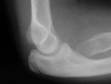
Fractures of the capitellum are rare. The complete capitellar fracture pattern was first described in the 19th century (1853) by Hahn and Steinthal; the eponym for this fracture pattern includes their names. Later, Kocher and Lorenz described an additional variation of this fracture pattern; a classification system includes their names.
Because of the rarity of capitellar fractures, controversies exist regarding the most appropriate treatment.[1] The fracture fragment is intra-articular and requires treatment and reduction to reestablish normal elbow motion. Difficulty arises from the varying size of the fracture fragment and from the varying amount of suitable subchondral bone that is present to achieve stable fixation and to allow early elbow motion. Failure of adequate intervention may result in an incongruous joint, as well as in stiffness, instability, and chronic pain.
For patient education resources, see the First Aid and Injuries Center, as well as Broken Arm and Broken Elbow.
NextFractures of the capitellum occur in the coronal plane. Separating the capitellum from the lateral column, capitellar factures are the result of shear forces from a fall onto the outstretched hand or of a fall directly onto the elbow. The capitellum is susceptible to shear forces because its center of rotation is 12-15 mm anterior to the humeral shaft.
Capitellar fractures may be associated with radial head fractures and posterior dislocations of the elbow. Other associated injuries include the disruption of the medial (ulnar) collateral ligament, the interosseous membrane, and the distal radioulnar joint.
Capitellar fractures account for 0.5-1% of all elbow fractures and 6% of all distal humeral fractures.[2] Capitellar fractures are seen with greater frequency in females than in males; this is thought to be secondary to a greater carrying angle and an increased possibility of osteoporosis in females. In 20% of patients with capitellar fractures, radial head fractures also are found.[3]
Capitellar fractures do not occur in children younger than 10 years. Because of the cartilaginous composition of the capitellum in children, a similar injury in a child would be a supracondylar or lateral condylar fracture.
Although some authors have advocated fragment excision, a study by Grantham et al demonstrated unsatisfactory results at 5-year follow-up.[4] The greatest complaint was stiffness and instability. In the same study, more favorable results were seen with open reduction and internal fixation (ORIF).
McKee et al also demonstrated improved results with early ORIF, along with early motion.[5] A 125º flexion-extension arc was achieved.
Despite the presence of greater flexion contractures at the time of follow-up in elbows with type IV fractures or fractures with an ipsilateral radial head fracture, good to excellent outcomes with functional ulnohumeral motion can be achieved following internal fixation of these complex fractures.[6]
Nonoperative and operative management of isolated capitellar fractures leads to satisfactory clinical outcomes as determined by postoperative range of motion (ROM), improvements in pain, and a return to previous levels of function.[7] No statistical difference in outcomes was observed between patients undergoing operative management and those undergoing closed reduction and immobilization
Ashwood et al presented the results of treatment of capitellar fractures in 26 patients who were followed prospectively and treated within a week of the injury.[8] According to the Mayo Elbow Performance Index (MEPI), nine patients had excellent results, nine had good results, and eight had fair results. Poorer outcomes were associated with posterior comminution of the humerus requiring more extensive procedures. All patients were able to return to work within 6 months, but 6 changed work roles from manual to administrative work.
Ruchelsman et al evaluated clinical, radiographic, and functional outcomes after ORIF of capitellar fractures in 16 patients.[6] Extensile lateral exposure and articular fixation with buried cannulated variable-pitch headless compression screws was performed at a mean of 10 days after injury. Injuries consisted of six type I, two type III, and eight type IV fractures according to the Bryan and Morrey classification. Supplemental minifragment screws were used in four of the type IV fractures and in one of the type III fractures. Mean ulnohumeral motion was 123º (range, 70º-150º).
Functional arc-of-elbow motion was achieved in 16 patients, and all patients had full forearm rotation.[6] Mean MEPI score showed nine excellent results, six good results, and one fair result. Patients with type IV fractures had greater magnitude of flexion contracture, reduced terminal flexion, and reduced net ulnohumeral arc.
Guitton et al studied 30 partial articular fractures involving the capitellum and trochlea.[9] One or more subsequent surgical procedures were required in 18 patients (67%), eight of which were for surgical complications. Routine removal of implants occurred in 15 patients. In addition to the fracture of the distal part of the humerus, four patients had an elbow dislocation; three had a fracture of the olecranon or the proximal part of the ulna; and two had a fracture of the radial head. The majority of capitellar fractures in this study turned out to be complex fractures of the articular surface involving both the capitellum and the trochlea.
Clinical Presentation
Copyright © www.orthopaedics.win Bone Health All Rights Reserved