One of the major thrusts in spine surgery today is to develop minimally invasive procedures. By definition, minimally invasive surgery utilizes small skin incisions, minimizes the damaging effects of large muscle retraction, and attempts to leave the body as naturally intact as it was prior to surgery. The goal is to achieve rapid recovery, lessen post-operative pain, and leave cosmetically satisfying incisional scars.
Post-operative Incisions Following Minimally Invasive Procedure
The purpose of this article is to introduce a remarkable new technology that enables surgeons to accurately implant spinal screws and rods in fusion surgery using computer technology and minimally invasive technique.
Only a small percentage of patients suffering from chronic back and leg pain will require fusion surgery. The common indications for a fusion procedure of the low back (lumbar spine) may include slippage of the spine (spondylolisthesis), recurrent disc herniation, chronic degenerative disc disease, traumatic fracture, or other forms of spinal instability. In fusion surgery, surgeons employ bone grafts and often stabilize the spine using screws and rods.
Traditionally, stabilizing screws and rods are placed on the spine through an “open” approach. This means there is a standard incision, which is typically up and down in the middle of the back. The large bands of muscles in the back are stripped free from their attachments to the spine and retracted off to each side. This allows for excellent visualization of the spine and easy access to the bones for implantation of the hardware. The downside of “open” surgery is that there can be considerable back pain from the muscle retraction, and the muscles develop some degree of permanent scar formation and damage as a result of the necessary retraction.
“Open” Approach
Though the goal of implanting instrumentation (screws, rods) to stabilize the spine remain the same, minimally-invasive techniques use the power of computer-assisted image guidance to allow the surgeon to “see” the spine through the skin without making a large incision. A special type of x-ray machine called a fluoroscope has been integrated with computer technology to enable surgeons. This system is referred to as “virtual fluoroscopy” (FluoroNav™). FluoroNav™ allows the surgeon to locate and navigate the spine using familiar x-images in real time but with greater power and accuracy, and with only a fraction of the usual radiation exposure. When combined with new instruments designed to be placed through small incisions, this form of computer-assisted, image-guided surgery becomes quite powerful.
The pedicle is a strong portion of the spinal vertebral bone that connects the front of the spine to the back of the spine. There is one pedicle on each side of each vertebral bone. Thus, placing a screw into the pedicle bone of the vertebral body proves to be a very strong way of purchasing or fixating the spine. Screws of all various designs used in this way are called pedicle screws. Once pedicle screws are placed at several levels of the spine, a rod can connect them all together on each side, giving the spine considerable extra strength.
One popular form of pedicle screws is made of titanium metal and has a head that can rotate to accommodate various anatomical conditions and positions. This threaded type of screw that can rotate is called polyaxial (can move about many axes). A new device called the SEXTANT™ allows for placement of polyaxial screws and pre-cut rods to be delivered to the spine through the skin. Open procedures with long skin incisions can be avoided. The SEXTANT™ device is what delivers the polyaxial screws along a trajectory provided by FluoroNav™ (computer-assisted image guidance).
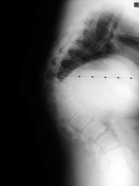
Pedicle Screws Implanted
SEXTANT™ Pedicle Screw Implantation: Integration of Titanium Screw Technology, Minimally Invasive Spinal Approaches, and Computer Technology
When a screw is placed through a very small incision in the skin, it is called a percutaneous procedure. Using the SEXTANT™ device, the surgeon navigates the spine using images provided by the FluoroNav™ device and places screws straight through the skin, fat, muscle and finally into bone. The screws are placed without disturbing the natural connections of muscle and tendons to bone. The SEXTANT™ device prepares a path through these tissues by bluntly dissecting through and creating a path. Then, a specially cut rod is passed through this path right into the heads of the screws. Using virtual fluoroscopy (FluoroNav™), each step of the procedure is “visualized” but without radiation exposure. The screw-rod connection is then tightened, and the SEXTANT™ device is removed. The patient is left with a stronger spine thanks to the stabilizing screws and rods, but all that is visible on the outside are a few small incisions the size of a fingernail.
Benefits
The initial experience with SEXTANT™ and FluoroNav™ have proven that it is a safe, effective way to implant spinal instrumentation that achieves the same result as open surgical placement of rods and screws. The following, however, are clearly emerging as benefits of percutaneous procedures over the conventional open operation:
Conclusion
Post-operative studies have clearly proven the efficacy, accuracy and reliability of the SEXTANT™ . Surgeons and patients nationwide are recognizing the power of computer-assisted, image-guided surgery done with the minimally invasive approach.
Copyright © www.orthopaedics.win Bone Health All Rights Reserved