The skeletal dysplasias are a heterogeneous group of disorders characterized by intrinsic abnormalities in the growth and/or remodeling of cartilage and bone. These dysplasias affect the skull, spine, and extremities in varying degrees.[1] They frequently cause a disproportionately short stature (dwarfism); the standing height falls below the third percentile for age. Achondroplasia is the most common type of short-limb disproportionate dwarfism.
The term achondroplasia, implying absent cartilage formation, was first used by Parrot in 1878.[2] Although the word achondroplasia is inaccurate from a histopathologic perspective, its use is universal and accepted by the International Working Group on Constitutional Diseases of the Bone.[3, 4, 5]
NextThe bony skeleton is divided into two parts: the axial skeleton and the appendicular skeleton. The axial skeleton is the central core unit, consisting of the skull, vertebrae, ribs, and sternum. The appendicular skeleton comprises the bones of the extremities. For more information about the relevant anatomy, see Skeletal System Anatomy in Children and Toddlers, Skeletal System Anatomy in Adults, and Osteology (Bone Anatomy).
Dwarfing conditions are frequently referred to as short-limb or short-trunk types, according to whether the trunk or limbs are more extensively involved. Achondroplasia, hypochondroplasia, and metaphyseal chondrodysplasias are considered short-limb dwarfing conditions. These patients' sitting height is within normal range. Additional terms used to describe the segment of the limb with the greatest involvement are rhizomelic (proximal), mesomelic (middle), and acromelic (distal). In achondroplasia, the extremity involvement is rhizomelic, with the arms and thighs more severely involved than the forearms, legs, hands, and feet.[6]
The primary defect found in patients with achondroplasia is abnormal endochondral ossification. Periosteal and intramembranous ossification is normal. Tubular bones are short and broad, reflecting normal periosteal growth. The iliac crest apophyses (appositional growth) are normal, giving rise to large, square iliac wings. The growth of the triradiate cartilage (endochondral growth) is abnormal, giving rise to horizontal acetabular roofs. Thus, the patterns of defect help to explain many of the observed clinical and radiographic characteristics of achondroplasia.
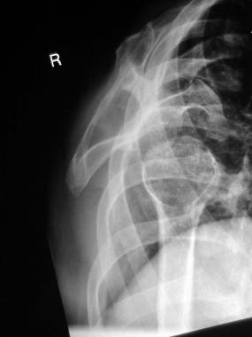
The characteristic features of achondroplasia are apparent at birth. The diagnosis is made on the basis of physical examination and radiographic findings.
A single gene mapped to the short arm of chromosome 4 (band 4p16.3) is responsible for achondroplasia and is transmitted as an autosomal dominant trait.[7]
At least 80% of cases result from a random new mutation. In sporadic cases, a paternal age older than 36 years is common. Most parents are of average size and have no family history of a dwarfing condition. The risk of the parents producing a second affected child is almost negligible. Reports have estimated that there is a 1 in 443 risk of recurrence of achondroplasia in the siblings of an affected child with unaffected parents. This is because of gonadal mosaicism in the parents. Average-sized siblings have no increased risk of producing a child with achondroplasia.
When both parents have achondroplasia, 50% of their offspring are heterozygous and affected, 25% are homozygous, which is ordinarily fatal in the first few months of life, and 25% are unaffected. When one parent has achondroplasia, the chance of transmitting this gene to each child is 50%.
Fibroblast growth factors (FGFs) are structurally related proteins associated with cell growth, migration, wound healing, and angiogenesis. At the cellular level, their function is mediated by transmembrane tyrosine kinase receptors, known as FGF receptors (FGFRs).[8]
Mutation in FGFR3 is responsible for achondroplasia, hypochondroplasia, and thanatophoric dysplasia.[8] The primary function of FGFR3 is to limit osteogenesis. Mutation causes enhancement in its function of limiting endochondral ossification. Mutation in FGFR3 in achondroplasia is due to transition of guanine to adenine (G to A) at nucleotide 1138 of complimentary DNA.
Two reports exist of achondroplasia associated with Down syndrome.[9] On the basis of the current birth rate, the calculated risk of association is 1 case in every 8 years in the United States.
Approximately 10,000 individuals are estimated to have achondroplasia in the United States. Worldwide, achondroplasia is the most common skeletal dysplasia, affecting about 1 in every 40,000 children. (This number varies, depending on the source.) About 80% of all "little people" have achondroplasia. Approximately 150,000 persons have achondroplasia worldwide. The worldwide population of little people is approximately 190,000.
Achondroplasia occurs with equal frequency in males and females. (It is inherited in an autosomal dominant manner.) Achondroplasia occurs in all the races with equal frequency.
The standardized mortality ratio is increased for all age groups by a factor of 2.27 over that of the general population.[10] In children younger than 4 years, death most commonly occurs as a consequence of brainstem compression, which causes sudden death. In individuals aged 5-24 years, central nervous system (CNS) and respiratory abnormalities are the common causes of death. In persons aged 25-54 years, cardiovascular problems are the most frequent causes of death.
Morbidity associated with achondroplasia may include the following:
An important resource for individuals with short stature is the Little People of America. This is a national organization that addresses the social, physical, and medical needs of its constituency. It holds annual regional and national conventions. Philosophically, these organizations emphasize the positive aspects of their members' abilities and lives rather than viewing the issue of short stature as a disability.
The Dwarf Athletic Association of America is a member of the US Olympic Committee that promotes athletic participation for individuals with short stature.
When addressing height issues in patients with short stature, the term "less than average" should be used.
Clinical Presentation
Copyright © www.orthopaedics.win Bone Health All Rights Reserved