Fractures of the hand are among the most common fractures of the skeletal system. Most of these fractures are acquired in the workplace or as a result of crush injuries, falls, or sports injuries.
The majority of these injuries can be managed nonoperatively, but certain fractures, such as intra-articular fractures, open fractures, unstable fractures, and displaced or angulated fractures, may require surgical correction with Kirschner wires (K-wires), plates, or screws. If these fractures are not treated properly, malunited fractures may result, leading to considerable loss of function and cosmetic disfigurement. Malunited fractures involving the joint surfaces can ultimately lead to posttraumatic osteoarthritis.
Malunion may be defined as healing of a fracture in an abnormal (nonanatomic) position. In the hand, it presents a combination of a functional problem with an aesthetic problem. It should be kept in mind, however, that the existence of a hand malunion does not necessarily mean that there is a dysfunctional hand or finger, because this is not often the case.
Management of malunion of hand fractures (see Treatment) is more complex than management of malunion of fractures elsewhere in the skeleton.[1, 2, 3, 4] Good hand function depends on joint mobility, sensibility, good skin coverage, adequate vascularity, and the gliding of a complex flexor and extensor tendon mechanism. Preexisting problems related to any of these factors may limit the usefulness of the digit, and surgical intervention can cause additional scarring and dysfunction. Consequently, management of hand malunion is predicated on a careful analysis of the risks and benefits of surgical intervention and on the functional goals and the likelihood that the operation can achieve them.[1]
NextMalunion is the most common bony complication of phalangeal fractures. The following four patterns of deformity are recognized:
Malrotation usually is seen after oblique or spiral fractures of the proximal and middle phalanges. The best method of assessing malrotation is to ask the patient to make a fist and look for digital overlap.
In adults with proximal phalangeal fractures, volar angulation exceeding 25-30° may result in pseudoclawing. This deformity makes using the hand awkward and can result in a fixed flexion contracture of the proximal interphalangeal (PIP) joint. The appearance may be aesthetically unacceptable.
Lateral angulation and malrotation often occur concomitantly. If correction is considered, the components of the deformity must be carefully identified.
Shortening may occur after a comminuted fracture is allowed to heal in a collapsed fashion or after a long spiral fracture.
In malunion of metacarpal neck fractures, sunken knuckle may be the clinical presentation. It is more of a cosmetic problem than a functional problem. In metacarpal shaft malunion, tendon imbalance and intrinsic contracture of the PIP joint may occur; however, function may still be preserved.
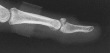
Intra-articular malunion occurs when intra-articular anatomy is not restored. Unreduced condylar fractures that extend into the PIP joint may produce pain, angulatory deformity, limited mobility, and, ultimately, degenerative arthritis.
Regarding metacarpal fractures, malunion can follow a transverse fracture, which results in dorsal angulation in the sagittal plane. Compensatory hyperextension (pseudoclawing) at the metacarpophalangeal (MCP) joint can result. Malunion after a spiral or oblique fracture results in malrotation. In patients with second and third metacarpal fractures, dorsal angulation is bothersome both cosmetically (pseudoclawing) and functionally. The prominent metacarpal head in the palm can be painful when the individual grips.
Rotational malunion of metacarpal fractures results in overlapping of the affected finger over an adjacent finger. The cosmetic deformity is often marked, and the grip is often impaired.
After crushing injuries or open fractures, shortening and associated problems of the soft tissue (eg, tendon adhesions, poor skin coverage, neurologic deficit) may occur.
Malunion most commonly affects the scaphoid among the carpal bones. Malalignment after union is evident as carpal collapse initially, and is reflected later in direct measurements of intrascaphoid alignment. The lateral appearance on radiographs shows the typical humpback scaphoid, which describes a deformity resulting from flexion angulation between the proximal and distal poles. Scaphoid malunion can alter carpal mechanics, leading to pain, weakness, limited motion, and degenerative arthritis.
Some authors have performed cadaveric studies to find the relationship between metacarpal shortening and extension of the MCP joint or the PIP joint. Strauch et al observed that for every 2 mm shortening of the metacarpal, there was a 7° lag in extension of the MCP joint.[5] However, this is not seen clinically, because of the ability of the MCP joint to hyperextend.
Vahey et al found that for every 1 mm of shortening of the proximal phalanx, there was a 12° lag in the PIP joint extension.[6] They also found that there was a linear relation between proximal phalanx shortening and PIP joint extensor lag and that increased angulation of the phalangeal fracture led to increased lag in extension of the PIP joint.
Malunion of hand fractures may result from inadequate treatment or failure of treatment. Accurate anatomic restoration may not be the goal of nonoperative treatment or even certain operative treatments for hand fractures. Hence, inaccurate anatomic restoration after treatment may not be considered evidence of inadequate treatment.
Most fractures of the hand bones occur in young, active adults who are involved in many various occupational and sporting activities. If these fractures are not managed carefully, they may result in malunion. This may lead to loss of function due to malalignment, malrotation, or shortening, which may result in decreased and disordered motion of fingers and poor outcomes.
The frequency of malunited fractures may be high in the hands, but few of these malunions require treatment. This is especially true with malunion of metacarpal neck fractures of the little fingers, which seldom produce deformity or interfere with function and therefore typically require no treatment.[2] In a study by Tubiana, out of 10,000 hand injuries, only 30 malunions required treatment.[7]
If treated carefully, with adherence to the principles described (see Treatment), most phalangeal and metacarpal malunions heal without clinically significant complications. Complications may include recurrence of deformity, neurovascular complications, or both.
Some patients may develop stiffness and decreased mobility. Most poor results are documented in elderly patients (>65 years) and in patients with crush injuries or extensive soft-tissue contractures. A combination of these factors increases the risk of compromised results. Proper selection of implants and quick rehabilitation may improve the prognosis.
In a study comparing 218 little-finger metacarpal shaft and neck fractures treated nonoperatively (with no attempt at fracture reduction) with 44 treated operatively with fracture reduction and fixation, severity of palmar angular deformity did not affect the outcome of nonoperatively treated fractures.[2] There were no differences in outcome between operatively treated and nonoperatively treated metacarpal neck fractures; and Disabilities of the Arm, Shoulder, and Hand (DASH) scores and aesthetic outcomes were better for metacarpal shaft fractures treated nonoperatively than for those treated operatively.
Potenza et al reported clinical and radiographic medium-term results for 24 fingers in 20 patients who underwent surgery for posttraumatic malunion of the proximal phalanx.[8] In all cases, corrective osteoclasia or osteotomy was done at the malunion site, followed by miniplate and screw fixation or by screw fixation only. Corrective osteoclasia was performed when malalignment was addressed within 6 weeks after injury. Two patients who had two fractures underwent additional surgery to improve function and range of motion.
Final follow-up occurred at a mean of 24 months after corrective surgery.[8] Good or excellent clinical and radiographic results were obtained for all patients. An improvement in grip strength was demonstrated by all patients. The mean score on the DASH symptom scale was 5 points. The researchers concluded that osteotomy in situ, in conjunction with stabilization by miniplates or screws, is effective for correcting posttraumatic malunions of the proximal phalanges of the fingers.
Clinical Presentation
Copyright © www.orthopaedics.win Bone Health All Rights Reserved