Limping in children is fairly common. The list of potential etiologies is long and varied and ranges from the benign to the life-threatening.[1, 2, 3, 4] Patients most often present to their primary care physician or an emergency department for initial evaluation. An understanding of normal and abnormal gait is critical.[5]
Once a gait abnormality has been identified, a thorough history and physical examination should be undertaken to narrow the differential diagnosis. The history should focus on the characteristic of the pain itself, as well as the presence of systemic or constitutional symptoms. Physical examination can help localize the etiology. Patients often have tenderness or loss of range of motion at the site of pathology. Further laboratory and radiographic studies are obtained based on the findings of the history and physical examination.
Well-defined treatments exist for most causes and vary with the severity of the disease process or injury. Fortunately, most children respond well to therapy and resume walking normally without sequelae.
Various pathologies are responsible for limping in children. At times, differentiating normal developmental changes from disease states presents a difficult dilemma. Establishing a diagnosis can be quite challenging, and these patients often require assessment by more than one physician in more than one visit. Aggressively pursuing the source of a child's limp at the first visit is essential to ensure optimal outcomes in the most patients.
Appropriate and early referral to an orthopedic specialist can benefit selected patients tremendously. However, in many instances, a conscientious physician can accurately assess and treat many of the conditions discussed in this article. Regardless, successful treatment of the child presenting with a limp demands sound clinical judgment, judicious ancillary testing, understanding of the possible differential diagnoses, and knowledge of therapeutic options.
For patient education resources, see Juvenile Rheumatoid Arthritis.
NextNormal synchronous gait develops in the first 3 years of life and requires the child to meet numerous anatomic and physiologic milestones. At age 1 year, many children can walk without support. By age 18 months, most children walk, and many can run. Coordination with reciprocal arm swing develops by age 2 years. Appropriate gait is attained with adequate musculoskeletal development of the lower back, pelvis, and lower extremities. Normal neurologic growth is mandatory for coordination and balance. Myelinization in a cephalocaudal pattern may explain the lack of complete control over those muscles that are integral to adult gait.
Finer adjustments to the gait pattern may not occur until the child is age 8-10 years, when normal adult gait pattern is attained. Adult gait patterns assume coordination of the following five key maneuvers:
However, for the purposes of this article, a more simplistic view of normal gait is described.
Normal gait begins with the stance phase, which is the weightbearing phase; it starts with the heel-strike. As the foot begins to plantarflex, the end of this phase culminates with the toe-off. The swing phase begins with toe-off and ends with the heel-strike. During the swing phase, coordinated gait requires forward rotation and tilting of the pelvis, as well as stability of the lumbar spine and abdomen. A limp or deviation from the normal expected walking pattern may be due to pain, weakness, or a structural abnormality.
Abnomalities of gait include the following:
A thorough history should be obtained from the child, if possible, and the parents. Parents' input is integral to obtaining an accurate perception of the complaint. A limp may originate from disorders affecting the abdomen, genitourinary tract, back, pelvis, hip, knee, foot, or other areas of the body.
Specific characteristics of the pain should be queried. A mnemonic that may be helpful in this regard is OPQRST, defined as follows:
Additionally, the presence of systemic or constitutional symptoms is very important. The presence of fever, weight loss, night sweats, anorexia, and/or general malaise may indicate a more serious etiology.[6, 7, 8, 9, 10]
The child's previous medical and surgical history should be ascertained as well as recent travel, immunizations, sick contacts, and a history of the child's development.
A complete physical examination is critical. The ultimate goals of the examination are to identify the type of limp (eg, antalgic, Trendelenburg, spastic) and localize the site of pathology.
The physical examination should begin with an overall assessment of the child, including his or her vital signs. Ideally, the child should be observed while barefoot and minimally clothed for assessment of stance and gait.
The child should be observed walking and running, if possible. Take the child into an open area to observe several gait cycles to elicit the gait abnormality and anatomic location. Running often accentuates subtle abnormalities. Range of motion of the individual joints should be observed. Identification of the specific type of nonantalgic gait should be made, if possible. The differential of a child with a Trendelenburg gait may be different from that of the child with an equinus gait.
The abdomen, pelvis, back, and extremities of the supine or sitting child should be inspected and palpated. Inspection and palpation are best accomplished when the child is sitting comfortably in the mother's lap. Areas of erythema, swelling, and warmth should be noted. Each digit and joint should be examined for motion and ligamentous stability.[11] Special attention should be given to the hips.
Neurovascular status, including strength, sensation, and reflexes, can also be assessed while the child is sitting or supine. Measure and compare lower-extremity lengths.
Laboratory assessment often begins with a complete blood count (CBC) and differential white blood cell (WBC) count, erythrocyte sedimentation rate (ESR), and possibly a C-reactive protein (CRP) level.[12, 13] These laboratory studies should be obtained when an infectious, inflammatory, or neoplastic etiology is suspected.
Blood cultures often are obtained when the index of suspicion is high for septic arthritis or osteomyelitis.
Common tests, such as electrolyte levels (eg, calcium), coagulation studies, and uric acid levels (to assess for gout), are occasionally necessary.
Sickle cell tests, Lyme disease titers, immunologic lab studies (eg, lupus antibodies, anti–double stranded DNA, rheumatoid factor, human leukocyte antigen, creatine kinase) are occassionally indicated when a specific etiology is supected.
A study by Dubois-Ferrière et al found that in the initial workup of limping children with suspected transient synovitis of the hip, many of the common diagnostic tests are unnecessary; according to the authors, routine assessment need include only the WBC count, the CRP level, the ESR, and hip radiography and ultrasonography.[14]
In 2012, the American College of Radiology presented its biannual Appropriateness Criteria, consisting of evidence-based guidelines to rate the appropriateness of imaging and treatment procedures for specific clinical conditions. The guidelines were based on an analysis of the medical literature in peer-reviewed journals and the application of a well-established consensus methodology.[15]
Standard radiography is the initial imaging study of choice in the limping child. (See the images below.) Orthogonal images of the area of concern should be obtained. Because of the high incidence of referred pain, radiographs of the hips are routinely obtained. Orthogonal images of the hip should consist of anteroposterior and frog-leg lateral views, with the exception of a suspected slipped capital femoral epiphysis, in which case a frog-leg lateral position could lead to displacement and a true lateral should be obtained. In very young children or those whose physical examination does not reveal a focal source of pain, radiographs of the entire lower extremity should be obtained. As many as 20% of children have unsuspected fractures.[16, 17]
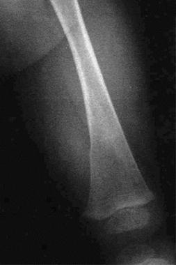 Distal femur fracture due to child abuse.
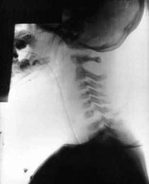
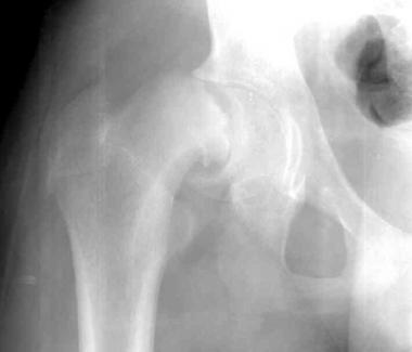 Slipped capital femoral epiphysis.
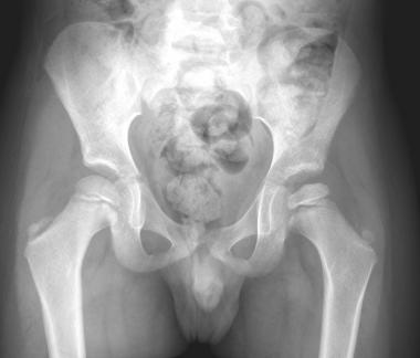 A 5-year-old boy with Legg-Calve-Perthes Disease.
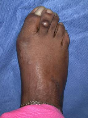
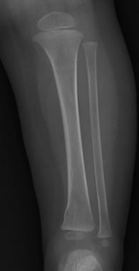 A 10-month-old boy with left distal tibia buckle fracture.
A study from Scotland using a protocol that radiographically assessed only the affected anatomic site had good success diagnosing the cause of limp.[18]
Ultrasonography is also often used.[19, 20] It is effective in diagnosing a hip effusion; however, it is unable to differentiate a sterile from a purulent effusion. In the patient with a high concern for septic arthritis, ultrasonography can be useful for guiding aspiration.
Bone scintigraphy is frequently used when the history and physical examination and other imaging studies do not identify the source of injury or pathology. Bone scintigraphy with technetium-99m highlights areas of increased bone metabolism[21] and may help to identify occult fractures,[22] osteomyelitis,[23] stress fractures, tumor or metastatic disease, and Legg-Calvé-Perthes disease.[24]
One study found bone scanning to be a beneficial initial test after radiography for children aged 2-11 years who had no clear etiology for foot pain, whether bilateral or unilateral.[25] In a small group of patients (49), bone scans aided in both diagnosis and treatment for those children who had not received an advanced study such as computed tomography (CT) or magnetic resonance imaging (MRI). For some patients, obtaining a bone scan may be a good alternative when the diagnosis is not clear after history, physical examination, and plain radiography.
CT is effective for abdominal and pelvic pathology (eg, sacroiliac trauma) and bony pathology of the hip, knee, spine, and foot.
MRI is the study of choice for soft-tissue pathology. MRI is also very useful in diagnosing stress fractures, osteomyelitis, malignancy, and early Legg-Calvé-Perthes disease.
For patients with suspected septic arthritis, the joint or joints are aspirated.[26, 27] The technique for aspiration varies according to the surgeon's preference.
Synovial fluid analysis should include cell count and differential WBC count, glucose, culture and Gram stain, and crystal examination.
Mucin clot formation suggests a noninfectious etiology, as bacteria break down the normal hyaluronic acid found in synovial fluid.
Bone aspirates can be assessed with Gram stain and culture for identification of the bacteria that is causing the osteomyelitis.
Knowing the child's age is imperative when considering the cause of a limp. A brief summary of common disease states in different age groups is listed in Table 1 below.
Table 1. Common Causes of Limping in Children (Open Table in a new window)
Age Common Types of Causes of Limping Common Causes of Limping 0-4 y Developmental Hip dysplasiaMost pathologic processes of the foot are nontraumatic in toddlers. Examining the children's shoes and feet is imperative. Possible etiologies causing a limp include the following.
Avascular necrosis of the navicular head (Köhler disease)[28] or second metatarsal (Freiberg infarction) may occur.
Microtrauma to the foot can result in stress fractures. Bone scans may be necessary for identification.
Ingrown toenails may cause limping. Standard incision and drainage and appropriate nail care are recommended.
Osteomyelitis may require both medical and surgical therapies, depending on the area of the foot involved and the extent of infection.[29] Subperiosteal abscess and failure to improve with medical management are indications for surgical treatment.
Puncture wounds can be managed conservatively with good local wound care and close follow-up. Patients who return with a wound infection or persistent pain should be evaluated for osteomyelitis, particularly that caused by Pseudomonas aeruginosa.
Toddler's fracture, a spiral fracture of the distal one third of the tibia, is often stable and incomplete and may be the result of a trivial injury, which is often not witnessed.
Torus fractures resulting from impaction, spiral fractures resulting from torsional injury, and greenstick fractures resulting from direct trauma occur commonly in children.[30]
Physeal injuries are more commonly seen in the older child following a rotational injury.
The knee and patellofemoral joints are common sites of pain, particularly in the older child and adolescent.
Sports-related injuries and overuse syndromes as well as infection may affect the knee area.
A popliteal, or Baker, cyst may rupture. These cysts are found along the inferomedial aspect of the knee.
Osteochondritis dissecans represents separation of articular cartilage, usually of the distal femur from underlying bone.
Osgood-Schlatter disease, affecting the patellar tendon insertion into the tibial tubercle, usually occurs in adolescents.
Patellofemoral syndrome is often the result of tracking abnormalities of the patella or patellofemoral instability. Jumping, running, and climbing may all be affected as the articular surface of the patella becomes inflamed.
Knee effusions may be related to overuse, trauma, or systemic disease. Thorough history taking, laboratory screening, and arthrocentesis with appropriate referral are suggested. The differential diagnoses include juvenile arthritis, lupus erythematosus, and infectious arthritis (including Lyme disease). A knee effusion after injury, especially in the adolescent athlete, may signal ligamentous injury.
Lyme disease can present in a variety of ways; synovitis, pauciarticular arthritis, and polyarticular arthritis may occur.
Bloody effusions may be associated with trauma. Possible traumatic causes include distal femur and proximal tibia fractures, tibial spine avulsions, patella fractures, and, rarely, knee dislocations.
Trauma, metabolic diseases, neoplasia, congenital deformities, and infection affect the thigh and femur.
Pathologic fractures related to tumors (benign or malignant), metabolic bone disease, or child abuse should be suspected with femoral shaft fractures. A fracture through a unicameral bone cyst in the proximal femur is not uncommon.
Metaphyseal beak fractures and rotational injuries that cause spiral deformities suggest possible child abuse.
Contusions of the thigh musculature are common in patients of all ages but may occasionally be debilitating and serious.
The distal femoral physis grows faster than any other physis in the body. According to Phemister's law, malignant and benign bone tumors occur in regions of the most rapid growth. Malignant tumors, including osteogenic sarcoma and Ewing sarcoma, generally produce more cortical destruction and periosteal reaction on radiographs than do benign lesions.
Osteomyelitis is usually due to hematogenous seeding, but it may also be the result of direct inoculation. Pain and swelling at the distal femur may represent osteomyelitis. In half of cases, the causative organism is Staphylococcus aureus. No organism is identified in up to one quarter of cases. Examine radiographs of the affected bone for abnormalities within the soft-tissue planes, cortical destruction, and periosteal elevation or thickening. Magnetic resonance imaging (MRI) or technetium-99m bone scanning may assist in localization.
Developmental dysplasia of the hip is thought to be due, in part, to laxity of the hip joint capsule, prenatal and postnatal position, and genetic and environmental factors.[31] When recognized and treated early, most dislocatable hips stabilize in the first 4-6 weeks of life and develop without sequelae. On occasion, the femoral head remains dislocated, thereby precluding normal development.
Useful physical examination tests include the Ortolani maneuver, Barlow test, and Galeazzi sign. Ultrasonography or plain radiography may confirm the findings. Patients with late or undiagnosed dysplasia often present with a waddling gait or a painful limp.
In children, trauma to the hip is more likely to result in a dislocation than a fracture. Prompt reduction of the dislocated hip usually results in few complications.
Hip fractures in children are associated with numerous complications, including avascular necrosis, premature closure of the epiphyseal plate, coxa vara, and nonunion.
Older children may experience avulsion of muscular attachments to the pelvis or proximal femur. Avulsion of the iliopsoas from the lesser trochanter or disruption from the ischial apophysis can occur (especially after sporting-event injuries).
Differentiating between transient synovitis and septic arthritis of the hip can be challenging. Both present with the leg held in flexion, abduction, and external rotation, representing the position of largest capsular volume. Clinically, most children with septic arthritis are toxic and present with fever, anorexia, and joint pain. Features of septic arthritis include hematogenous spread and purulent effusion. Articular destruction is due to proteolytic enzymes, most commonly S aureus. The hip, knee, and ankle are most commonly affected.
Ultrasonography of the hip has been shown to be effective to assess for osteomyelitis, septic arthritis, transient synovitis, Legg-Calvé-Perthes disease, and developmental dysplasia of the hip.[32, 33, 34, 35, 36]
Kocher et al proposed a clinical prediction algorithm to help differentiate between transient synovitis and septic arthritis of the hip.[37] The probability of septic arthritis was predicted on the basis of the following four factors:
This algorithm has not been prospectively tested. Kocher et al recommended diagnostic arthrocentesis in patients with two to four positive factors and close observation for patients with no predictors. Patients with only one positive variable had a 5% or less chance of having septic arthritis.
An elevated C-reactive protein level has been shown to be a good indicator of septic arthritis and should be included as part of the evaluation.
On synovial fluid analysis, a WBC count higher than 50,000/μL with a predominance of polymorphonuclear (PMN) cells favors a diagnosis of septic arthritis, which is a surgical emergency.[38] Transient synovitis is generally associated with lower counts and fewer PMN cells. Gram stain and culture help guide antibiotic therapy.
Legg-Calvé-Perthes disease typically affects boys in the first decade. Avascular necrosis of the capital femoral epiphysis results in remodeling of the femoral head and acetabulum over 1-3 years. Flattening of the femoral head may be a late finding. Acute presentations are associated with synovitis of the hip.[39, 40]
Slipped capital femoral epiphysis usually affects large-for-age adolescents.[34] It presents with a limp associated with hip pain or referred pain to the thigh or knee.[6] Delayed diagnosis is not uncommon due to referred symptoms. Most cases are unilateral, but bilateral involvement is possible.[35] The second slip often presents 6-18 months later. Anteroposterior pelvic radiographs may reveal subtle widening of the physis, although the frog lateral projection may be more helpful at identifying subtle slips. A Klein line, drawn along the superior femoral neck, may intersect less or none of the femoral head on the affected side.
Hip pathology often presents with thigh or knee pain.
Infection, rheumatologic disease, microtrauma, and neoplasia of the spine, pelvis, or abdomen may result in a limp. In general, these disease processes are more common in the older child or adolescent.
Juvenile ankylosing spondylitis and rheumatoid arthritis may cause back pain.
Neoplastic diseases affecting the pelvis, spine, and abdomen include neuroblastoma, leukemia, osteogenic sarcoma, and renal tumors.
Overuse syndromes are common in active adolescents.
Spondylolysis due to a stress fracture of the pars interarticularis is found most commonly at L5.
Herniated nucleus pulposus in the older child or adolescent presents in a similar fashion as in adults but is much less common.
Discitis, inflammation of the intervertebral disc, may be infectious or, less likely, may be sterile. MRI may be necessary to identify the pathology.
Abdominopelvic lesions, which manifest solely as a limp, are uncommon. Such pathology often involves the retroperitoneum and includes appendicitis, psoas abscess, retroperitoneal hematoma, and renal/ureteral inflammation.
Pelvic entities include osteomyelitis, genitourinary infections, and tumors or hernias.
Skin and lymphatic diseases in the inguinal region may cause pain, swelling, and a limp.
Copyright © www.orthopaedics.win Bone Health All Rights Reserved