A unicameral bone cyst (UBC) is a common, benign, fluid-filled lesion found almost exclusively in children. Much has been written about the diagnosis and management of these lesions, and evidence of a variety of successful treatment strategies can be found in the literature.
The orthopedic entity called a UBC is not believed to be a new phenomenon. Lagier et al identified a UBC in the femur from the remains of a child from medieval times.[1] Virchow also recognized such bone cysts in humans in the late 1870s.[2]
In 1942, Henry Jaffe and Louis Lichtenstein published their classic paper concerning solitary UBCs.[3] In their article, the authors emphasized the distinctiveness of UBC as follows:
"Solitary unicameral bone cyst is a lesion sui generis. It bears no relation whatever to giant cell tumor of bone, and in particular it does not represent a cystic-healing phase of this tumor. Nor is it to be linked with enchondroma, fibroma or focus of fibrous dysplasia of bone that has undergone partial or extensive cystic degeneration. Further, it should not be regarded as representing cystic expression of osteitis fibrosa, since to throw it into this wastebasket category (one which to us is also meaningless) is to obliterate its distinctiveness. Correspondingly, solitary unicameral bone cyst ought no longer to be classed as an expression of localized fibrocystic disease of bone or localized fibrous osteodystrophy—likewise blanket designations dating from a more primitive era of bone pathology."
Despite abundant clinical confidence in managing these lesions, many basic questions remain concerning the etiology and pathophysiology of UBCs.
This article offers a comprehensive review of the present state of knowledge of a UBC, highlighting aspects of the pathophysiology, the clinical presentation, and the most commonly used treatment strategies.
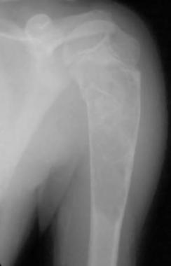
NextA UBC is a benign, fluid-filled, radiolucent lesion that may appear in virtually any bone, but typically, it is found in either the proximal humerus or proximal femur. (See the images below.) A UBC often leads to thinning of adjacent areas of bone, such that fracture or pain from microfracture may occur.
 Large proximal humeral unicameral bone cyst demonstrates early cortical healing following pathologic fracture.
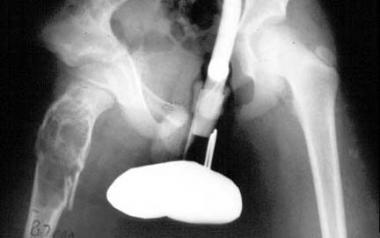 Large unicameral bone cyst of pelvis. Pathologic fracture is depicted. Note extension of cyst into region of proximal femoral physis.
When UBCs are immediately adjacent to a growth plate, they are referred to as active cysts, and when they have achieved some distance from the growth plate, they are considered to be latent cysts. This distinction has been used in the past; it was believed to have prognostic significance. A UBC usually presents as a unifocal (one bone) problem, affecting patients who are skeletally immature.
The rarity of the lesion in adults supports a hypothesis of spontaneous resolution. In the absence of fracture through the cyst (or impending fracture), UBCs are asymptomatic. They are, at times, found serendipitously when radiographs are taken for other reasons. In the absence of symptoms and in the absence of mechanical compromise of the involved bone (eg, extensive cortical thinning), no treatment may be necessary other than observation.
However, treatment should be strongly considered for lesions that have resulted in a fracture or marked weakening of bone. Some evidence exists that spontaneous healing of a UBC may occur following fracture. Such healing occurs in only a minority of cases. Growth disturbance secondary to a UBC is also a concern.[4]
At least two case reports exist in which a chondrosarcoma was found to arise within the same area as a previous histologically proven UBC.[5] In a separate case, an 8-year-old boy was reported to have sustained a pathologic fracture of the distal fibula that was believed to have resulted from a Ewing sarcoma infiltrating a UBC.[6]
The precise relation between such rare instances of apparent malignant transformation and the thousands (if not millions) of UBCs that have not demonstrated such behavior remains unclear. At any rate, a UBC is not considered to be a malignant or premalignant lesion; accordingly, routine biopsy or other treatment of asymptomatic and nonproblematic lesions based on a patient's or family's fear of cancer should not be undertaken.
A UBC occurs most frequently in children aged 5-15 years (average age, ~9 years).[7, 8] Many authors consider cysts that present in the first decade of life to be more aggressive.[7, 8] A UBC affects males approximately twice as often as females. These lesions constitute approximately 3% of all bone tumors.
A UBC probably represents the third or fourth most common benign bone tumor that the orthopedic surgeon confronts (osteochondromas are commonly considered to be the most frequently encountered benign bone tumors in children, followed by fibromas and/or fibrous cortical defects). The lesion may occur in conjunction with other benign bone tumors, such as a nonossifying fibroma.[9]
By far, the most common location for the lesion is the proximal humerus, followed by the proximal femur. The proximal humerus and femur together account for nearly 90% of all UBC sites.[7, 8] However, virtually any bone may be affected, with the calcaneus being one of these notable alternative locations.[10, 11, 12, 13, 14, 15]
The specific etiology of a UBC has not been elucidated. Many theories have been proposed. One commonly quoted theory was proposed in 1960 by Cohen,[16] who studied the cyst fluid from six children undergoing treatment for UBCs and found four to resemble plasma and two to resemble blood. Cohen proposed that the principal etiologic factor is blockage of the drainage of interstitial fluid in a rapidly growing and rapidly remodeling area of cancellous bone.
Chigira et al studied the internal pressure of 7 patients with UBCs and found them to be higher (2-7 mm Hg range) as compared to the contralateral normal bone marrow pressures.[17] The arterial oxygen tension (PaO 2 ) in the fluid from these same cysts was found to be impressively lower than that in venous or arterial samples taken at the same time. These authors suggested that venous obstruction within the bone appears to be a likely cause of such simple bone cysts.
Such vascular theories have been supported by other authors.[18] Mirra et al suggested that a UBC represents an area of a congenital rest of synovial tissue and was able to demonstrate both synovial type A (macrophagelike) and type B (fibroblastlike) cells in the lining of such cysts.[19] This description resembles that of an intraosseous synovial cyst. Yu et al also demonstrated how methylprednisolone influences the cellular physiology of synovial cells in culture, thus establishing a theoretic basis for steroid injection treatments for a UBC.[20]
Shindel et al reported increased prostaglandin E2 levels in the cyst fluid from 7 of their patients and theorized that this may help explain the beneficial effect of steroid injection of such lesions.[21] Gerasimov led a group of Russian researchers who stressed that the fluid from UBCs possesses increased lysosomal enzyme activity regardless of the UBCs' status as active or latent.[22] These authors emphasized the role such enzymatic activity might play in permanent corrosion of the cyst cavity, as well as increasing osmotic pressure within the cyst.
High levels of cytotoxic oxygen free radicals have also been found in the fluid from UBCs.[23] Such free radicals are not only cytotoxic; they might be generated during the ischemic state following blockage of interstitial fluid drainage from UBCs. The Japanese researchers suggested that such oxygen scavengers may contribute to the bone destruction associated with UBCs. Reproduction of these results in other centers has not yet occurred.
In the past several years, a group of Brazilian researchers have reported specific genetic abnormalities in a pediatric patient with a UBC of his right distal femur. Vayego et al made their first report in 1996.[24] Cytogenetic analysis of the resected cyst initially demonstrated complex aberrations of chromosomes 4, 6, 8, 12, 16, and 21. Further study of the same patient (following bone cyst recurrence) later revealed specific mutations associated with amino acid substitutions (arginine for tryptophan, arginine for serine).[25]
More study in this area clearly is indicated, and the potential for future gene-based therapies is seemingly apparent.
Komiya and Inoue carried out a longitudinal study (with serial radiographs over 6 years) that documented the development of a UBC over time.[26] Initially, a small erosive lesion of the endosteal humeral metaphysis appeared, and over time, the lesion progressively enlarged into a typical UBC. The lesion analyzed by these authors was somewhat unusual, in that it was located in the distal humerus. In addition, the lesion appeared following notation of a previous UBC in the proximal aspect of the same bone.
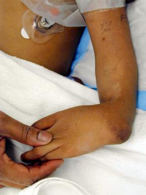
Most patients with a UBC present to the orthopedic surgeon after sustaining a pathologic fracture. Such fractures most commonly involve either the proximal humerus or the proximal femur. The events leading up to the fracture may range from fairly trivial (eg, throwing a ball) to more substantial (eg, a hard fall while playing soccer). As with all patients who have sustained a fracture, a careful physical examination of the patient should include efforts to exclude the possibility of an open fracture and a neurocirculatory insult.
In other instances, patients may present to emergency department physicians, their primary care physicians, or orthopedic surgeons for other reasons, and radiographs obtained in the workup of other complaints may identify asymptomatic UBCs. Such incidental diagnosis of "a bone tumor" may be quite disconcerting to the child's parents and family. Random bone tumor discussions with such a child's family are contraindicated. Medical personnel who eagerly deliver well-intentioned but inaccurate discussions of bone tumors often needlessly terrify families.
In either scenario, a review of the patient's past history, as well as their family's past history relative to fractures, rheumatologic conditions, bone tumors, endocrine disease, and cancer, is appropriate. Physical examination of the patient should also include a screening examination of the axial skeleton and the uninvolved extremities. Any other identified abnormalities may require further plain radiographs.[27] Palpation of major lymph node areas (eg, the axillary and inguinal fields) is also appropriate; infection and malignancy are part of the differential diagnosis.
The decision to pursue surgical intervention in patients with UBCs is a highly individualized one. An asymptomatic lesion with satisfactory maintenance of cortical thickness may require only observation. A lesion with precarious cortical thinning (with or without insufficient pain) may demand surgical intervention. In addition, factors such as an upper extremity (lower stress) versus a lower extremity (higher stress) and younger children (more amenable to cast immobilization) versus older adolescents (less amenable to cast immobilization) may strongly influence surgical decisions. Simple treatment of the pathologic fracture may result in cyst resolution in up to 25% of cases.[28]
Some authors have suggested the use of a cyst index aimed at predicting the future risk of a pathologic fracture. Andre Kaelin and Dean MacEwen discussed this concept and defined their cyst index as the area of the UBC measured via its widest dimensions divided by the diameter of the diaphysis of the same bone.[29] Based on their statistical analysis of 57 patients with UBCs, these authors recommended mainly observation for humeral cysts with an index of less than 4 and for femoral cysts with an index of less than 3.5.[29] When either of these thresholds was exceeded, stronger consideration regarding surgical intervention was believed to be appropriate.
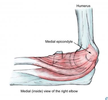
The anatomy that is relevant to UBCs is mainly that of the proximal humerus and proximal femur. Percutaneous approaches to the proximal humerus require the surgeon to avoid injury to the biceps tendon as well as the axillary nerve as it innervates the deltoid musculature. The standard deltopectoral approach is the most common open surgical approach for proximal humeral lesions.
Key points of this approach include preservation of the cephalic vein as well as careful medial retraction of the conjoined tendon (coracobrachialis and short head of the biceps) to avoid injuring the musculocutaneous nerve. Dissection in the region of the bicipital groove should be minimized; such dissection may injure the anterolateral ascending branch of the anterior humeral circumflex artery, which provides the bulk of the blood supply for the humeral head.
The main contraindication for surgical treatment of a UBC is a patient who otherwise meets indications for surgery but is unable to tolerate anesthesia. Another relative contraindication for surgery is a patient with a small asymptomatic latent cyst with a low likelihood of a pathologic fracture.
Workup
Copyright © www.orthopaedics.win Bone Health All Rights Reserved