Fibrous dysplasia (a term first suggested by Lichtenstein and Jaffe in 1942[1] ) of bone is a nonheritable disease in which abnormal tissue develops in place of normal bone. Abnormalities may involve a single bone (monostotic form; 70% of cases) or many bones (polyostotic form; 30% of cases). The polyostotic form is occasionally associated with precocious puberty, fibrous dysplasia, and cafe-au-lait skin lesions (McCune-Albright syndrome, Albright syndrome) or with myxomas of skeletal muscle (Mazabraud syndrome).[2, 3, 4, 5, 6]
The etiology of this abnormal growth process is related to a mutation in the gene that encodes the subunit of a stimulatory G protein (Gsα) located on chromosome 20.[7, 8] As a consequence of this mutation, a substitution occurs in which the cysteine or the histidine—amino acids of the genomic DNA in the osteoblastic cells—is replaced by arginine.[9]
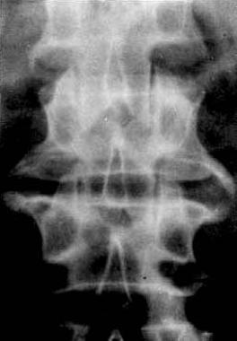
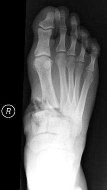
Fibrous dysplasia lesions are characterized by woven ossified tissue and extensive marrow fibrosis. Mechanical quality of bones is decreased. As a consequence of this bone fragility, patients have an increased risk of fracture. Incidence of fractures is around 50% of cases.[10] This risk of fractures or bone deformity is higher in the long bones, such as the femur, tibia, and humerus, but all the bones can be affected.
Pain is a common symptom of patients with fibrous dysplasia. Patients also have an increased risk of malignant tumors such as osteosarcoma, fibrosarcoma, chondrosarcoma, and malignant fibrohistiocytoma.[11] The incidence of this risk has been evaluated to be reduced to 1%.[11, 12] This risk is higher in patients with the polyostotic form, or McCune-Albright syndrome.[12]
NextAs a consequence of the mutation of GNAS1, there is a substitution in which cysteine or histidine, amino acids of the genomic DNA in the osteoblastic cells, is replaced by another amino acid, arginine.[9]
Osteoblastic cells expressing this mutation have a higher DNA synthesis than normal bone cells. The growth of these cells is faster, leading to an inappropriate differentiation of mesenchymal cells. At the molecular level, intracellular cyclic adenosine monophosphate (cAMP) levels are increased and osteocalcin is decreased.[13] Osteocalcin is a late marker of osteoblast differentiation. Involved bone cells are immature. They fail to produce normal amounts of collagen or to orientate appropriately to the lines of mechanical stress.
Fibrous dysplasia is caused by the sporadic mutation of the GNAS1 gene, which encodes the alpha subunit of the stimulatory G protein (G1) located on chromosome 20q13.2-13.3 of the osteoblastic cells.[8] Although the mutation is known, the actual pathways that lead to abnormal osteoblast differentiation and function are just beginning to be understood.
The consequence of this mutation is an inappropriate cell differentiation resulting in a disorganized fibrotic bone matrix. Cancellous bone maintenance is perturbed, and bone undergoing physiologic remodeling is replaced by an abnormal proliferation of fibrous tissue.
The extent and pattern of disease depend on the stage of development and the location at which the mutation occurs. All the bones can be affected.
Fibrous dysplasia accounts for about 5% of all benign bone tumors.[8] The monostotic form is more common than the polyostotic form. Because many patients are asymptomatic, the true incidence of this disorder is unknown. Usually, fibrous dysplasia presents clinically in children and adolescents, with a median onset age of 8 years. Most cases manifest themselves before the age of 30 years. Males are affected more often than females, except in McCune-Albright syndrome, in which females are affected more often than males.
Monostotic fibrous dysplasia is active while it is growing but often becomes inactive after puberty. It may reactivate during pregnancy. Polyostotic disease typically remains active throughout life.
Unless malignant transformation develops, fibrous dysplasia is not a life-threatening disease. The lesions tend to stabilize as skeletal maturity is reached. The majority of the monostotic cases have a good evolution regardless of treatment. Polyostotic disease tends to have a poorer prognosis.[14] Polyostotic lesions are very often associated with one or more fractures.[15] Malignant transformation develops in a minority of patients (<0.5%).
The recurrence rate for fibrous dysplasia has been reported to be 21% after curettage and grafting, but if patients are monitored for many years, the rate is probably closer to 100%.
Clinical Presentation
Copyright © www.orthopaedics.win Bone Health All Rights Reserved