Synovial chondromatosis is a rare condition in which foci of cartilage develop in the synovial membrane of joints, bursae, or tendon sheaths as a result of metaplasia of the subsynovial connective tissue. These ectopic foci of cartilage can result in painful joint effusions and, on the generation of loose bodies, mechanical symptoms.[1, 2, 3, 4, 5]
Controversies surrounding the diagnosis and treatment of synovial chondromatosis are few. Fukuhara et al identified elevated levels of chondrocalcin in the synovial fluid of patients with synovial chondromatosis.[6] They suggest that this finding may assist in diagnosing the disease, though the utility of this test does not justify its expense at this point. Similarly, levels of interleukin (IL)-6 and vascular endothelial growth factor (VEGF)-A in synovial fluid may someday have clinical utility, but at present, they remain of primarily academic interest.
The pain, swelling, and mechanical symptoms of synovial chondromatosis and its generation of loose bodies have historically been treated with surgery. Since the mid-1950s, the technical strategy has included open arthrotomy with removal of loose bodies and synovectomy. Controversy exists regarding the utility of synovectomy. Some authors prefer simple loose-body removal and limited excision of involved synovium only. Open synovectomy and loose-body excision remain acceptable treatments. Sufficient evidence supports arthroscopic removal of loose bodies with limited synovectomy as first-line surgical therapy.
NextSynovium lines the interior surface of diarthrodial joints and is composed of vessel-rich fronds lined by synoviocytes. Ectopic cartilage bodies in the synovium and loose in the joint must be removed. The need for total synovectomy, including removal of normal areas of synovium, is questionable. Total synovectomy can lead to clonally significant stiffness after surgery, with reported rates of up to 43% when the procedure is performed by means of open arthrotomy.
Dorfmann et al and Coolican and Dandy reported low recurrence rates after arthroscopic treatment of the knee and no postoperative stiffness with simple excision of loose bodies.[1, 4, 7] In current practice, most authors agree that arthroscopic removal of loose bodies for mechanical symptoms is the best surgical strategy.
Primary synovial chondromatosis appears to occur in the following three phases, as described by Milgram in 1977[8] :
Synovial chondromatosis is considered a benign process associated with an extremely low risk of malignancy. Case reports have described the coexistence of chondrosarcoma and synovial chondromatosis, sparking debate as to whether the chondromatosis is a cause or the result of chondrosarcoma. Only a few case reports have documented malignant degeneration of synovial chondromatosis.[9, 10, 11, 12, 13]
A review of 155 cases of primary synovial chondromatosis by McCarthy et al identified only four cases of aggressive behavior and chondrosarcomalike histology (one in the elbow and three in the hip).[14] All of the tumors behaved as locally aggressive neoplasms; none gave rise to metastases.
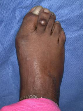
Involvement is typically monoarticular, with the large joints being most frequently affected. The knee joint is involved in 60-70% of cases; the shoulder, elbow, and hip are the next most frequently involved joints. Reports have described involvement of multiple other joints and locations, including the following[1, 2, 3, 15, 16, 17, 18] :
Some histochemical evidence shows low proliferation activity in the synovial loose bodies. In tissue samples taken from diseased joints, Ki-67, a general marker of proliferative activity, has been found to be minimally expressed in the loose bodies, but with increased expression in the synovial membrane. High levels of bone morphogenic protein (BMP)-2 and BMP-4 mRNA have also been isolated from free bodies and synovial tissue. It is thought that these may promote cartilaginous and osteogenic metaplasia. Additionally, elevated levels of IL-6, aggrecan, and VEGF-A have been found in cases of synovial chondromatosis of the temporomandibular joint.[19, 20, 21]
Synovial chondromatosis occurs as either a primary or secondary form. Although the molecular basis is still unclear, high levels of BMP-2 and BMP-4 have been isolated from diseased synovium and free bodies.[5] These growth factors may be involved in the pathologic metaplasia observed in synovial chondromatosis.
This form is described as the presence of ectopic cartilage in synovial tissue and as loose bodies in the joint cavity with or without calcification (osteochondromatosis) and without an identifiable joint pathology. Whether this represents synovial metaplasia or a true neoplasia is unclear. The true etiology remains unknown, but most authorities favor the metaplastic theory.
Secondary synovial chondromatosis is more common than primary disease. It occurs in the setting of preexistent osteoarthritis, rheumatoid arthritis, osteonecrosis, osteochondritis dissecans, neuropathic osteoarthropathy, tuberculosis, or osteochondral fractures. Free chondral or osteochondral fragments formed by underlying disease implant into the synovium and induce metaplastic cartilage around them. The free bodies are typically larger and of more varied sizes than those seen in primary disease.[22]
A rare cause of secondary synovial chondromatosis has been documented in an extra-articular bursal space following resection of a benign osteochondroma. It was thought that some chondral debris from the surgical excision may have remained after the index procedure.[23]
The exact prevalence of synvolial chondromatosis is unknown, but the disorder is rare worldwide. Most reported series indicate a male-to-female ratio of 2:1. In addition, most cases are reported in patients aged 20-40 years; only a few case reports have described the condition occurring in children.[22]
In current practice, most authors agree that arthroscopic removal of loose bodies for mechanical symptoms is the best surgical treatment. This strategy minimizes postoperative stiffness associated with open procedures and successfully accomplishes synovectomy and loose body removal. Longer follow-up has validated the success described in early arthroscopic reports.
Dorfmann et al demonstrated that arthroscopic loose-body excision was successful in 25 (78%) of 32 knees.[24] Most failures were simple recurrences requiring repeat arthroscopic surgery. Four of 24 patients with prolonged functional impairment had substantial tibiofemoral arthritis at the initial time of treatment. At 2-year follow-up, Samson et al treated 11 of 13 patients with arthroscopic treatment. Of this group, six good and six very good outcomes were achieved, with only two patients requiring arthroscopic reoperation.[25]
Synovial chondromatosis may lead to articular damage and subsequent development of osteoarthritis. Ackerman et al retrospectively examined the outcome of joint arthroplasty applied in the setting of severe osteoarthritis in patients with synovial chondromatosis. With average follow-up of 10.8 years, all patients reported improvements in pain, range of motion (knees only), and functional scores. One of seven hips and one of four knees demonstrated radiographic reoccurrence.[26]
Evidence supporting arthroscopic treatment now is supported in other joints. Urbach et al evaluated arthroscopic treatment of synovial chondromatosis found in the shoulder. With follow-up between 4-9 years, 5 patients demonstrated very good clinical results. Of note, 2 patients were found to develop asymptomatic progression of disease.[27]
At a mean follow-up of 5.3 years, Lunn et al retrospectively evaluated 15 individuals treated by shoulder arthroscopic synovectomy, loose body removal, and selective bicep tenodesis for primary and secondary synovial chondromatosis. Nine patients demonstrated bicipital groove loose bodies, and of this subgroup, seven patients underwent open bicipital debridement and tenodesis. Disease reoccurrence occurred in only two patients at an average of 7.5 years.[28]
Less arthroscopic success has been demonstrated in the ankle. In 2008, Galat et al reviewed eight patients (average age, 37 years) who presented with ankle (n=6) or midfoot (n=2) synovial chondromatosis. In half of these patients, open synovectomy led to pain-free function without disease reoccurrence. Unfortunately, three patients ultimately underwent below-knee amputation for reoccurrence (n=1) or low-grade malignant transformation.[11] A single case report of successful arthroscopic treatment of synovial chondromatosis of the ankle joint has been reported.[29]
Synovial chondromatosis of the hip is uncommon. In 2006, Schoeniger et al reviewed eight patients with monoarticular synovial chondromatosis of the hip who had joint debridement and a modified total synovectomy performed through a surgical hip dislocation with a trochanteric flip osteotomy. With average follow-up of 6.5 years, two patients had symptomatic progression necessitating joint replacement. The remaining six patients demonstrated no disease reoccurrence, progression of osteoarthritis, and continued pain relief.[30]
In 2008, Boyer and Dorfmann reviewed 120 patients who underwent arthroscopic management for primary synovial chondromatosis of the hip. With average follow-up of 78.6 months, 37.8% of 111 patients required further open surgery, and 20.7% required additional arthroscopic surgery. Of this original population, 56.7% reported excellent or good outcome, but 19.8% eventually underwent hip joint replacement.[31]
Subsequently, Lee et al published their results from 1996 through 2008. Twenty-four patients with synovial chondromatosis of the hip underwent arthroscopic removal of loose bodies and synovectomy, with a mean follow-up of 41 months. The average patient was able to walk and bear weight 2 days after surgery. Four patients demonstrated worsening of their arthritis over the follow-up period, with one requiring conversion to a total hip arthroplasty. Harris hip scores improved from a preoperative average of 39 to a postoperative average of 82. Overall, 75% had a good or excellent results, with four patients demonstrating recurrent disease.[32]
Similarly, Marchie et al at Massachusetts General Hospital published their results from a series of 29 patients in 2011. They found a 43% good or excellent result at an average follow-up of 60 months, with five patients eventually undergoing conversion to a total hip arthroplasty at an average of 52 months. Of note, they found that imaging studies alone failed to establish a diagnosis in 48% of their patients, with definitive diagnosis being made at the time of arthroscopy.[33]
Ferro et al studied clinical presentation, associated pathology, and clinical outcomes in 23 patients who underwent arthroscopic surgery for synovial chondromatosis of the hip between 2005 and 2012.[34] All 23 experienced improvements in range of motion. The average Modified Harris Hip Score rose from a preoperative value of 62 to a postoperative value of 84.8; the Short Form (SF)-12 phsyical component summary rose from 41 to 53; and the Western Ontario and McMaster Osteoarthritis Index improved from 27.1 to 7.2. Median overall satisfaction was 9.5/10.
Given the results of these studies, arthroscopic management for synovial chondromatosis is appropriate for disease isolated to the knee, shoulder, and hip. For reasons that remain unclear, this success has yet to be evaluated and/or confirmed to ankle, elbow, and minor joints. Regardless, outcomes for patients with severe, symptomatic arthritis and secondary synovial chondromatosis seem to generate less predictable results.
Clinical Presentation
Copyright © www.orthopaedics.win Bone Health All Rights Reserved