Hand infections include superficial infections, infections of the nail, infections of the tendon and tendon sheath, infections of the deep spaces of the hand, septic arthritis, and osteomyelitis.[1] They can range from routine problems (treated with oral antibiotics, immobilization, and limited incision and drainage)[2] to catastrophic surgical emergencies (resulting in significant compromise of hand function). The purpose of this article is to provide a systematic approach to the diagnosis, evaluation, and treatment of hand infections.
Hand infections will continue to be a routine problem encountered by emergency physicians, primary care physicians, and hand specialists.[3] The clinician needs to be aware of the increasing incidence of infections with more virulent microorganisms.[4] Community-acquired infections with methicillin-resistant Staphylococcus aureus (MRSA) are encountered in nearly every area of the body, and the hand is no exception.[5, 6] In addition, with the growing number of cancer survivors, transplant patients, and patients living with HIV infection, the surgeon can anticipate treating more complex polymicrobial hand infections.
For patient education resources, see the Skin Conditions and Beauty Center, as well as Hand Injuries and Finger Infection.
NextA brief review of the most common hand infections by anatomic location follows.
Acute paronychia involves the soft tissue around the fingernail and usually results from the inoculation of bacteria (most commonly S aureus) into the paronychia tissue from nail trauma or nail manipulation.
Drain superficial abscesses with limited incision and drainage. Obtain cultures if possible. If the infection resulted from an ingrown nail, excision of the radial or ulnar one fourth to one half should be performed at the time of incision and drainage. Infections that involve the eponychial fold can be drained by elevating the eponychium, either sharply or with a freer or elevator. The patient should receive a course of oral antibiotics with good staphylococcal coverage (eg, IV cefazolin or oral cephalexin). In addition, the patient should soak the finger in antiseptic solution 2-3 times a day.
Chronic paronychia usually is caused by Candida albicans and occurs most commonly from chronic immersion in water (as in dishwashers), previous trauma, or nail defects. Treatment with topical antifungal agents and behavior modification is occasionally successful. Excision of a portion of the nail or removal of the entire nail may be necessary.
A felon is a subcutaneous abscess over the distal pulp of a digit or thumb. Felons usually result from a penetrating injury. The pulp contains multiple compartments separated by fibrous septa that make infections in this area complex.
Surgical drainage is necessary when an area of palpable fluctuance is present. Use of several incisions has been described for drainage. However, the preferred incision is radial or ulnar longitudinal. Incisions directly over the finger pad or tip are avoided. Subcutaneous septa should be broken up to drain all areas of infection, and the wound is left open. After drainage, warm antiseptic soaks and oral antibiotics are administered. The antibiotic is based on the nature of the infection. Parenteral antibiotics should be considered in patients with diabetes or in those who are immunocompromised. Persistent chronic paronychial infections may also require intravenous (IV) antibiotics.
Deep-space infections in the hands are possible. The two deep spaces in the palm are the midpalmar space and the thenar space. Infections in these areas usually result from injuries such as bites or puncture wounds. Such infections may cause cellulitis, fluctuance, or pain.
In addition, the second, third, and fourth web spaces are potential sites for infection. Web-space infections can spread from the palmar subfascial space in a dorsal direction, forming what is commonly referred to as a "collar button abscess." On examination, patients typically have pain, swelling, and fluctuance on the palmar or dorsal web-space surface.
Flexor tenosynovitis is a potentially devastating infection that can result in significant scarring of the flexor tendon sheath with resultant compromise in hand function.[7, 8] These infections usually are caused by a penetrating injury (eg, bite, puncture wound). In the early 1900s, Kanavel described the following tetrad of physical findings in patients with flexor tenosynovitis:
Flexor tenosynovitis may also occur without Kanavel signs, particularly in immunocompromised patients.
Kameyama et al studied stenosing flexor tenosynovitis (SFTS) in diabetic patients and nondiabetic patients to identify the relative frequency of multiple-digit involvement in these two populations. According to the authors, diabetic patients showed a significantly higher prevalence of multiple-digit SFTS than nondiabetic patients did, and limited joint mobility in diabetic patients was found to be closely associated with multiple-digit SFTS.[9]
In most cases, patients with flexor tenosynovitis require urgent incision and drainage of the flexor tendon sheath. Broad-spectrum antibiotic coverage against staphylococci is initiated after cultures are obtained. Then, culture-specific antibiotics are given.
Septic arthritis usually occurs as a sequela of open skeletal trauma or a bite wound. Patients with inflammatory arthritis are at increased risk for joint-space infections. Tenderness and swelling of the joint are signs of potential infection. Puncture wounds over the joint should suggest potential septic arthritis. The differential diagnosis includes gout, psoriatic arthritis flare, and systemic lupus erythematosus.
Staphylococci and streptococci are most commonly isolated in septic joint cultures. Arthrotomy is the preferred treatment, as opposed to joint aspiration. The latter can be used to aid diagnosis; however, arthrotomy is required for adequate drainage of the infection. The interphalangeal joints (proximal and distal) can be accessed through a dorsal or midaxial incision. The collateral ligaments often must be released to allow access to the joint capsule. The metacarpophalangeal joint can be accessed via a dorsal approach. A 10-day course of culture-specific antibiotics is required.
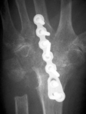
Osteomyelitis can occur from an acute event, such as a penetrating wound or open fracture, or as a late sequela of a fracture or other surgery. Patients with a history of diabetes or other immunocompromising conditions are at higher risk for osteomyelitis. Diagnosis of this condition is based on the signs seen with other infections: cellulitis, warmth, and tenderness. In addition, recurrent infections in the same location may be a sign of infection of the underlying bone. Laboratory studies and radiographs can assist in making the appropriate diagnosis (see Workup). The treatment consists of debridement of the devitalized bone, as well as antibiotics, usually a prolonged course of 6 weeks.
Herpetic whitlow is a viral infection that is caused by the herpes simplex virus and that may resemble a felon or paronychia.[10, 11] These infections usually occur in medical or dental personnel. History is an important clue to the diagnosis. The patient first notices pain, then erythema before the development of the herpetic vesicle.
Treatment of herpetic whitlow is nonoperative; therefore, differentiating these infections from bacterial felons and paronychia is important. The diagnosis can be confirmed by obtaining cultures of the vesicles. Overall, the infection has a self-limited course. Therapy consists of pain control. Topical antiviral agents have been recommended in patients who are immunocompromised. A 20% risk of reactivating the herpetic infection has been reported.
Hand infections usually result from an injury, most commonly a laceration or an animal bite.[12, 13] Most patients recall an inciting event that resulted in the inoculation of bacteria into the hand. Infections of the nail and of the nail folds can result from a nail deformity. One study found that the most common etiologies of wrist-joint infection were gout, pseudogout, and cellulitis; the incidence of septic arthritis was low.[14]
If the hand infection has been treated appropriately with measures such as eradication of the abscess and devitalized tissue, the risk of recurrence is minimal. Certain infections (eg, herpetic whitlow), however, have a 20% recurrence risk.
In patients with septic tenosynovitis of the hand, the presence of a subcutaneous abscess may be predictive of persistent or worsening disease and thus of the subsequent need for additional debridement after the first procedure.[15]
Clinical Presentation
Copyright © www.orthopaedics.win Bone Health All Rights Reserved