For descriptive purposes, as well as for operative considerations, forearm fractures are classified by location, being categorized as proximal, middle, or distal third fractures. The middle third of the radius stretches from the radial bow to the beginning of diaphyseal straightening. The ulna is relatively straight and can be divided using longitudinal dimensions alone. (See also Forearm Fractures, Distal-Third Forearm Fractures, and Forearm Fractures in Emergency Medicine, as well as Galeazzi Fracture and Monteggia Fracture.)
Treatment objectives for both-bone forearm fractures have remained relatively constant, with early extremity range of motion. To understand the management of forearm fractures, the idea of the forearm axis was created, combining the function and anatomy of the wrist, forearm, and elbow. The coordinated, independent function of the wrist, forearm, and elbow is necessary to place and orient the hand in space. Injury to any of these components can result in a significant deficit. The three basic stabilizers of the forearm are as follows:
For patient education resources, see the First Aid and Injuries Center, as well as Broken Arm.
NextUnlike fractures in infants and children, fractures of the adult forearm are unstable. Nonunions and malunions of both-bone forearm fractures are functionally and cosmetically limiting, with midshaft radius or ulna angulation substantially impeding forearm rotation.[1]
According to the AO (Arbeitsgemeinschaft für Osteosynthesefragen [Association for Osteosynthesis]) documentation center, forearm fractures accounted for 10-14% of all fractures between 1980 and 1996.
Middle third, or diaphyseal, forearm fractures commonly result from a direct blow or a fall from a height. Other causes include gunshot wounds, motor vehicle accidents, and pathologic bone fractures.
Patients with middle third forearm fractures present after an identifiable traumatic event. Multiple injuries to the musculoskeletal and associated systems frequently occur in conjunction with forearm fractures. In patients who sustain multiple traumas, any life-threatening injuries take priority in treatment and stabilization. Forearm fracture management objectives remain the same whether they are isolated or occur in the polytrauma setting, with early stabilization recommended.
Physical examination of the patient with a forearm fracture includes a close inspection of the skin to rule out puncture wounds and abrasions. Additional soft-tissue evaluation is performed to rule out compartment syndrome, which is associated with low- and high-velocity injuries. A careful neurologic and vascular examination is carried out to identify any deficits that were caused by the injury. Loss of posterior interosseous nerve (PIN) function in Monteggia fracture patterns has been well described. The PIN innervates the extensor musculature below the elbow, which functions to extend the digits.
Anterior interosseous nerve (AIN) palsy also may be present and is often overlooked because this finding has no sensory component. A division of the median nerve, the AIN arises from the posterior aspect of the median nerve, 5 cm distal to the medial humeral epicondyle, and passes between the heads of the pronator teres.
The AIN is primarily a motor nerve; injury to it can cause paralysis of the flexor pollicis longus (FPL) and flexor digitorum profundus (FDP-I) to the index finger, causing loss of pinch between the thumb and index finger. The AIN terminates in sensory fibers to the distal radioulnar, radiocarpal, intercarpal, and carpometacarpal joints. Palsy has been associated with internal fixation of forearm fractures, as well as with tight external dressings.[2]
Nonoperative treatment of middle third forearm fractures is reserved for isolated ulnar shaft fractures, better known as nightstick fractures. (See also Interventions for Isolated Diaphyseal Fractures of the Ulna in Adults.) Radiographs of the wrist and elbow must be obtained in isolated radius and ulna fractures to rule out Monteggia and Galeazzi injury patterns. These injuries are best treated surgically in the adult patient.
Both-bone middle third forearm fractures in adults are unstable injuries that lead to shortening and angulation. The goal of treatment is to achieve a stable anatomic reduction. The literature recommends open reduction and internal fixation (ORIF) for displaced fractures of the middle third of the forearm in adults to restore early forearm motion.
Precise anatomic reduction is necessary to reestablish the radial bow and proper interosseous space and therefore to maintain normal motion.[3] Even small amounts of malalignment may lead to a functional disability at the wrist and/or elbow.
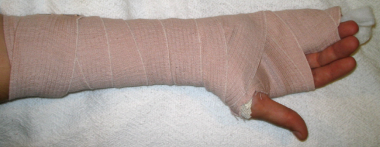
Nondisplaced both-bone middle third forearm fractures are rare in adults; when present, however, they may be treated in a long arm cast for 6-12 weeks with the elbow flexed to 90º and the wrist in neutral rotation. Careful weekly radiographic follow-up is required because these fractures may displace. If displacement occurs, ORIF is required to restore the normal anatomic relation of the radius and ulna.
All open forearm fractures require appropriate irrigation and debridement with subsequent surgical stabilization. Open fractures of grades I, II, IIIa, and (occasionally) IIIb are treated according to the same principles of closed fractures in association with meticulous debridement of the soft tissues. Results of debridement and immediate internal fixation of open fractures are comparable to those of surgical treatment of closed fractures.[4]
Open wound management is recommended, with repeated debridement as necessary. Primary closure of the extension incisions of the traumatic wound may be performed, and delayed wound closure may be performed once the soft tissues have declared themselves.
Forearm fractures accompanied by soft-tissue loss that results in an inability to cover plates may require other forms of stabilization. Temporary stabilization with an external fixator may be achieved while planning soft-tissue coverage with rotational or free flaps in conjunction with delayed (secondary) internal fixation (plating).
Most forearm fractures in children can be stabilized and treated by means of closed reduction and cast immobilization.[5] Occasionally, some pediatric forearm fractures can be unstable, leading to displacement, radioulnar angulation, rotational malalignment, and encroachment of the interosseous space.[1, 6] Angulation of greater than 10º results in loss of rotation in children older than 10 years and should be avoided.
Intramedullary (IM) stabilization was introduced in France in 1984 as an alternative to plate fixation in children, in an attempt to avoid extensive exposure and soft-tissue stripping.[7] Since then, IM fixation has gained widespread acceptance in the United States for surgical treatment of pediatric forearm fractures. Indications for IM fixation are as follows[8, 9, 10] :
The ulna is the stable unit about which the radius rotates. Force transmission is initiated at the wrist (the distal radioulnar joint) level and is translated longitudinally between the radius, ulna, and interosseous membrane, through the forearm axis, and to the elbow.
An applied compressive load travels along the proximal radius, transferring tension forces to the interosseous membrane, which transfers a compressive load to the proximal ulna.[11] This mechanism accounts for the inequality in the contact forces between the radius and ulna at the wrist and elbow. Fracture and/or dislocation of the forearm lead to disruption of this longitudinal relation and affects wrist, forearm, and elbow function.
To regain and reestablish function, the forearm joint must be reconstructed anatomically. Every effort should be made to maintain normal anatomic relations.
Contraindications to surgical treatment include life-threatening trauma conditions, which may delay or preclude surgical intervention. Rarely are patients so medically unstable that both-bone forearm fractures cannot be promptly treated by means of surgery.
Workup
Copyright © www.orthopaedics.win Bone Health All Rights Reserved