The term hallux varus describes a clinical condition of the foot characterized by medial deviation of the great toe relative to the first metatarsal ray. Hallux varus has variable degrees of severity, symptomatology, and etiology. Causes range from the most common iatrogenic postoperative variety[1] to idiopathic, rheumatic, and posttraumatic (tear of the hallux lateral collateral ligament) forms.
Fossilized evidence of modern humans in Africa from 3 million years ago reveals footprints that show hallux varus. Other imprints uncovered in northern Japan are dated to 2300 BC and also show varus alignment of the great toe. The later impact of shoe wear had a definite influence on alignment of the hallux.
Flexible hallux varus is a common finding in newborns and is a reflection of intrauterine positioning. It usually corrects to valgus in early childhood when walking begins.[2, 3] On the other hand, the normal 0-20º deviation that is seen in hallux valgus occurs after walking has begun in the child and after shoes have been introduced to the child's feet.
NextCadaveric biomechanical studies have revealed that the anatomic restraints to hallux varus, in descending order, are the lateral capsule, the adductor hallucis, and the lateral flexor brevis tendon.
Congenital hallux varus may be divided into primary and secondary pathologic deformities.[4] Primary hallux varus is a rare condition that is usually related to an overactive abductor hallucis. Secondary hallux varus is related to other congenital abnormalities, such as metatarsus adductus, great toe polydactyly, longitudinal epiphyseal bracket syndrome, and delta phalanx.
Primary dynamic infantile hallux varus is caused by medial insertion of the abductor tendon. Acquired adult hallux varus is described in inflammatory arthropathies, including rheumatoid and psoriatic arthritis. The mechanism of such arthropathies combines destruction of the articular surfaces by pannus, intrinsic muscular contracture, and distention of the joint capsule with subsequent laxity of the collateral ligaments.
Few reports exist of traumatic hallux varus following sports injuries. In the cases that have been reported, hallux varus occurred secondary to rupture of the lateral collateral ligament and conjoined tendon.
Spontaneous idiopathic hallux varus may be noted incidentally and is usually supple (see the images below).[5] An etiologic factor is not always demonstrable.
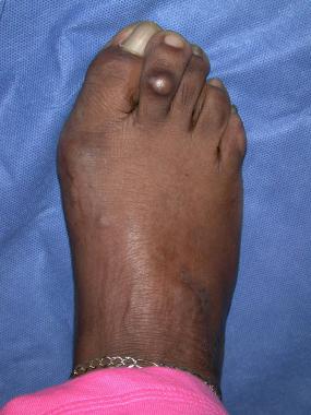
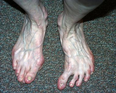 Clinical photo of idiopathic hallux varus of left foot.
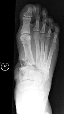
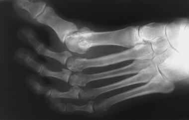 Anteroposterior radiograph of foot depicts idiopathic hallux varus.
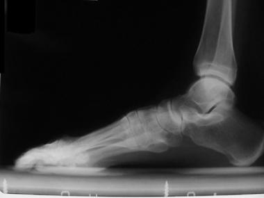 Lateral radiograph of foot shows idiopathic hallux varus.
The initial deforming force is likely overpull of the abductor tendon, which is related to medial insertion into the proximal phalanx; this may be influenced by an inflammatory process or by minor trauma. The imbalance leads to varus deformity and subsequent contracture of the medial capsule, decrease of the intermetatarsal (IM) angle, and medial subluxation of the flexor and extensor mechanisms.
Shoe wear tends to correct the varus deformity rather than exacerbate it, as it does for hallux valgus. Therefore, spontaneous idiopathic hallux varus may be more common than is reported.
The classic deformity of hallux varus occurs most frequently after a surgical procedure involving aggressive lateral soft-tissue releases, typically a distal soft-tissue or McBride[6] type of bunionectomy, but it can also be produced after Silver, Chevron, Mitchell, Keller, and Lapidus[7] procedures (see the images below).[8]
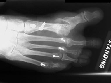 Anteroposterior radiograph of foot shows iatrogenic hallux varus following proximal osteotomy and distal soft-tissue realignment.
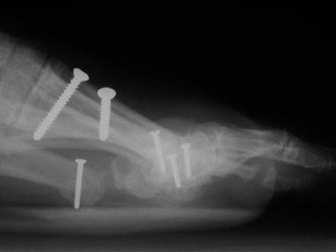 Lateral radiograph of foot depicts iatrogenic hallux varus following proximal osteotomy and distal soft-tissue realignment.
Classically, the deformity is characterized by the following:
This posture results from muscle imbalance that is brought about by the medial subluxation of the tibial sesamoid. Release or transfer of the adductor hallucis alone is not sufficient to produce dynamic hallux varus; however, when coupled with excision of the fibular sesamoid or transection of the lateral head of the flexor brevis tendon, hallux varus likely results. Other predisposing factors for hallux varus are a small IM angle and a round metatarsal head.
Flexion of the MTP joint is brought about by the flexor hallucis brevis through its pull on the sesamoid sling. If the fibular sesamoid is excised, the metatarsal may buttonhole through the defect and result in hyperextension and medial deviation of the MTP joint. Medial deviation is exacerbated when the adductor tendon is detached and nothing opposes the pull of the abductor hallucis.
Potential contributing factors include overplication of the medial capsular structures, medial displacement of the tibial sesamoid, overpull of the abductor hallucis against an incompetent lateral ligamentous complex, excessive resection of the medial eminence, and overcorrection with a postoperative dressing that holds the MTP joint in a varus position. Another cause of hallux varus is overcorrection of a proximal first metatarsal osteotomy, leading to a negative IM angle.
The incidence of iatrogenic postoperative hallux varus ranges from 0% for distal osteotomies without a lateral release to 15% for proximal osteotomies (specifically, the Lapidus procedure) with distal soft-tissue release.[9, 10] Most reports are of crescentic osteotomies, which have an overall varus rate of 10%. The incidence of idiopathic, congenital/infantile, traumatic, and otherwise acquired hallux varus, however, is unknown.
Surgery is aimed at improving the overall position of the hallux, not necessarily its motion. Preoperatively, surgeons must inform patients that further salvage procedures may be necessary and that most surgical procedures directed at correcting iatrogenic hallux varus are 60-80% effective.[11] In one series of patients treated with extensor hallucis brevis tenodesis, the American Orthopaedic Foot and Ankle Society (AOFAS) hallux MTP-IP score improved from 61 to 85.[12]
Clinical Presentation
Copyright © www.orthopaedics.win Bone Health All Rights Reserved