Shoulder arthrocentesis can be performed diagnostically for identification of the etiology of acute arthritis or therapeutically for drainage of an effusion. With the same technique, the joint can be injected therapeutically with corticosteroids, anesthetics, or both.
The practitioner performing the procedure should be familiar with the anatomy of the glenohumeral joint and surrounding structures in order to avoid complications. The glenohumeral joint of the shoulder is formed by the humeral head and the glenoid fossa of the scapula. It is bounded by the acromion. The subdeltoid bursa lies under the deltoid muscle and covers the lateral and superior aspect of the proximal humerus. The neurovascular bundle lies medially in the axilla.
Three glenohumeral ligaments exist, as follows:
The SGHL has a variable origin and inserts on the humerus near the lesser tubercle; it resists inferior translation of the humeral head in the adducted shoulder. The MGHL originates from the labrum and inserts on the humerus medial to the lesser tubercle; it resists inferior translation in the adducted and externally rotated shoulder. The IGHL originates from the labrum and the adjacent glenoid neck and inserts on the anatomic neck of the humerus; it resists humeral head anterior and posterior translation. (See Shoulder Joint Anatomy.)
Aspiration of the glenohumeral joint can be accomplished from an anterior or a posterior approach. The posterior approach allows the patient to be blinded from the procedure, and it mimics the approach used in arthroscopy of the joint.
Diagnostically, shoulder arthrocentesis is used in patients with shoulder pain for injection of anesthetic (with or without corticosteroid), to determine whether the glenohumeral joint is the source of the patient's pain. By permitting joint aspiration, arthrocentesis aids in the diagnosis of the underlying pathologic process through synovial fluid analysis.[1, 2]
Therapeutically, shoulder arthrocentesis can be used to provide pain relief and functional improvement in glenohumeral osteoarthritis, rheumatoid arthritis, or adhesive capsulitis (this may be performed through drainage of an effusion, septic joint, or hemarthrosis or through instillation of medication.)
Bacteremia, cellulitis of overlying skin, and adjacent osteomyelitis are often considered absolute contraindications because of the potential risk of seeding the joint with bacteria. In these situations, the procedure should be performed only if the clinician strongly suspects septic arthritis as the cause of overlying inflammatory changes, and then only after consultation with an orthopedist.
Relative contraindications include glenohumeral joint infection, chronic infection distant to injection site, allergy to injectate, diabetes mellitus, and uncontrolled coagulopathy.
NextLocal anesthesia is recommended. (See Local Anesthetic Agents, Infiltrative Administration.) After skin preparation and identification of the needle insertion site, use a 25-gauge needle to make a small skin wheal with local anesthetic into the subcutaneous tissue and then along the anticipated needle pathway.
Equipment for shoulder arthrocentesis consists of the following:
The patient should be seated in a comfortable position. For the anterior approach, rest the patient’s hand on his or her lap so that the shoulder is internally rotated. For the posterior approach, place the patient’s hand on the contralateral shoulder.
Steps in the performance of shoulder arthrocentesis are as follows:
Needle placement accuracy rates appear to be significantly higher for the posterior approach to the glenohumeral joint than for the anterior approach.[3] Accuracy rates are also higher when imaging is used in conjunction with injection and aspiration.
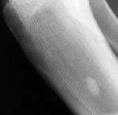
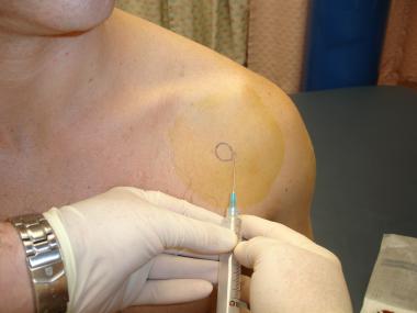
Palpate the coracoid process and the humeral head. As the arm is internally rotated, the joint space can be felt as a groove lateral to the coracoid process (see the image below).[4]
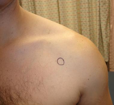 Circle represents coracoid process.
Insert the needle medial to the head of the humerus and just below the tip of the coracoid process (see the image below).
 Shoulder arthrocentesis. Insert needle medial to head of humerus and just below tip of coracoid process.
Direct the needle slightly laterally and superiorly into the scapulohumeral joint space (see the image below).[5]
Shoulder arthrocentesis. Direct needle slightly laterally and superiorly.
Insert the needle 1-2 cm inferior and medial to the posterior tip of the acromion. Direct the needle anteriorly and medially toward the coracoid.[6]
Complications are uncommon and often insignificant but include the following:
Copyright © www.orthopaedics.win Bone Health All Rights Reserved