Patellar dislocations are common, particularly in adolescent females and athletes. Patients usually present with an inability to extend an obviously deformed knee. A sizable effusion may also be seen. This injury may be due to direct trauma to the patella or to a valgus stress combined with flexion and external rotation.
The reported incidence of patellar dislocation is 5.8 per 100,000, but it may be as high as 29 per 100,000 in the adolescent population.[1] There are several varieties of patellar dislocation, as follows:
Several anatomic factors, including a lateralized tibial tubercle,[2] the tibial tuberosity – trochlear groove distance,[2] and the shape of the patella,[3] may increase the likelihood of lateral patellar dislocation.
Reduction of a lateral dislocation of the patella is a simple and safe procedure. Otherwise, an orthopedist should be consulted for these more uncommon types of dislocations.
NextNonsurgical reduction can be attempted on any lateral or medial dislocation of the patella. An immediate attempt at reduction should be made on any dislocation associated with vascular compromise of the distal extremity, though this is exceedingly uncommon in the setting of an isolated lateral patellar dislocation and should prompt further examination into concurrent injuries.
Some surgeons have advocated surgical rather than nonsurgical intervention to treat patellar dislocation, out of concern for possible recurrence. A Cochrane review by Smith et al found that although there was some evidence that appeared to favor surgical management of primary patellar dislocation, the quality of the currently available evidence was too poor to allow any firm conclusions to be made.[4]
A patellar dislocation associated with a fracture of the proximal tibia or distal femur should not be reduced in this manner. Osteochondral fractures may occur in the setting of patellar dislocation. Caution should be used in evaluating for a fracture, either by exam or by radiographs, prior to attempting reduction. Any superior, intercondylar, or horizontal dislocation should be examined by an orthopedic surgeon. Any dislocation with suspected locked osteophyte should be examined by an orthopedic surgeon.
Anesthesia is usually not required for this procedure, though some patients have significant anxiety and pain. Procedural sedation should be used as needed to maximize the patient's comfort during the reduction.
No equipment is needed for the reduction. A knee immobilizer, crutches, or both are needed for aftercare.
Place the patient supine or with the legs hanging off the side of a gurney (see the image below).
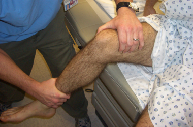 Positioning for lateral patellar reduction.
Explain the procedure, risks, and benefits to the patient. Lateral or medial reduction is a safe and technically simple procedure. Obtain informed consent for the reduction (and the procedural sedation if necessary). To optimize successful rehabilitation, educate the patient regarding aftercare.
Stand on the lateral side of the leg on which the patellar reduction is to be done. (See the video below.) Slightly flex the injured leg at the hip to decrease tension on the quadriceps muscles. Extend the knee while applying gentle, anteromedially directed force on the lateral patellar edge to lift the patella over the femoral condyle. For a medial dislocation, use the same technique, but stand medial to the dislocation and apply an anterolateral force. When reduction is complete, apply a knee immobilizer so that the knee is in full extension.
Conducting patellar reduction.Arrange a follow-up appointment for the patient with an orthopedic surgeon. Some patients with complete dislocation may require surgery to prevent recurrence.
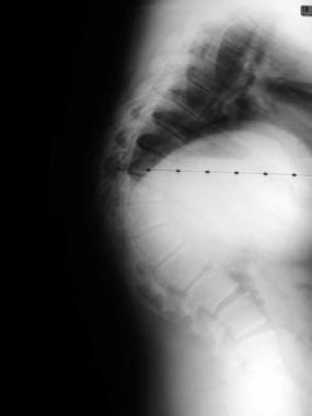
Obtain prereduction and postreduction radiographs to rule out any osteochondral fractures, if suspected based on mechanism of injury or by physical examination.
Computed tomography (CT) can detect small bony fragments that result from patellar dislocation. These fragments are often not seen on standard radiographs. CT should be considered in first-time dislocation patients and in dislocations that result from significant forces.[5]
The literature reports controversy regarding which patients should undergo operative repair of primary dislocations. Most patients do well with a short course of immobilization followed by physical therapy.[6, 7]
Medial patellofemoral ligament injury typically results from patellar dislocation[8] ; thus, follow up with an orthopedic surgeon is recommended for all patients with patellar dislocations.[9, 10, 11, 12, 13]
Osteochondral fractures are a very uncommon complication of reduction of a patellar dislocation. Related complications of the dislocation itself may include recurrent dislocations, degenerative arthritis, or osteochondral fractures.
Copyright © www.orthopaedics.win Bone Health All Rights Reserved