In 1703, William Musgrave first described a neuropathic joint as an arthralgia caused by venereal disease.[1] In 1868, Jean-Martin Charcot gave the first detailed description of the neuropathic aspect of this disease; hence, the condition is named after him.[2] Charcot noted this disease process as a complication of syphilis. Syphilis was believed to be the most common cause of Charcot arthropathy until 1936, when Jordan linked it to diabetes. Diabetes is now considered to be the most common etiology of Charcot arthropathy.
Sohn et al performed a retrospective study of patients with Charcot arthropathy to compare the risks of lower-extremity amputation in patients with Charcot arthropathy alone and those with diabetic foot ulcers. They found that Charcot arthropathy by itself does not pose a serious amputation risk, but amputation risk is multiplied in the presence of ulcer complications. The study showed that Charcot patients and ulcer patients had 4.1 and 4.7 amputations per 100 person-years, respectively. In patients younger than 65 years, amputation risk, compared with patients with Charcot arthropathy alone, was 7 times higher for patients with ulcer alone and 12 times higher for patients with Charcot and ulcer.[3]
Della Paola et al evaluated, as an alternative to amputation in patients with Charcot arthropathy, the use of surgical treatment of osteomyelitis of the midfoot or the ankle and stabilization with external fixation. In the study of 45 patients, 39 patients healed using emergent surgery to drain an acute infection while maintaining fixation for an average of 25.7 weeks; 2 patients were treated with intramedullary nails in follow-up surgery; and in 4 patients, infection could not be controlled and amputation was still necessary. The authors concluded that in select patients, external fixation may be an alternative to below-the-knee amputation.[4]
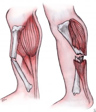
NextAlso called Charcot joint or neuropathic joint, Charcot arthropathy is a progressive condition of the musculoskeletal system that is characterized by joint dislocations, pathologic fractures, and debilitating deformities. This disorder results in progressive destruction of bone and soft tissues at weight-bearing joints; in its most severe form, it may cause significant disruption of the bony architecture. Charcot arthropathy can occur at any joint; however, it occurs most commonly in the lower extremity, at the foot and ankle.
The prevalence of Charcot arthropathy ranges from 0.1% to as high as 13% in specialized foot clinics. In patients with diabetes, the incidence of acute Charcot arthropathy of the foot and ankle ranges from 0.15-2.5%.
Epidemiologic studies do not distinguish between acute and postacute disease. Bilateral disease occurs in less than 10% of patients. Recurrence of disease occurs in less than 5% of patients. Some studies indicate that men and women are equally affected, while others report a 3:1 predilection for males.
Any condition that causes sensory or autonomic neuropathy can lead to a Charcot joint. Charcot arthropathy occurs as a complication of diabetes, syphilis, chronic alcoholism, leprosy, meningomyelocele, spinal cord injury, syringomyelia, renal dialysis, and congenital insensitivity to pain. Diabetes is considered to be the most common cause of Charcot arthropathy.[5] There is also evidence for a relationship between Charcot arthropathy and rheumatoid arthritis.[6]
The exact nature of Charcot arthropathy remains unknown, but the following major theories exist regarding the pathophysiology of this condition:
Charcot arthropathy most likely results from a combination of the processes described in the above theories. The autonomic neuropathy leads to abnormal bone formation, and the sensory neuropathy leads to an insensate joint that is susceptible to trauma. The development of abnormal bone with no ability to protect the joint results in gradual bone fracture and in the subluxation of the joint.
The clinical presentation of Charcot arthropathy can vary widely depending on the stage of the disease. Thus, symptoms can range from mild swelling and no deformity to moderate deformity with significant swelling.
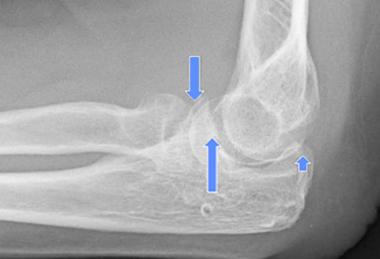
Acute Charcot arthropathy almost always presents with signs of inflammation. Profound unilateral swelling, an increase in local skin temperature (generally, an increase of 3-7 º above the nonaffected foot's skin temperature), erythema, joint effusion, and bone resorption in an insensate foot are present. These characteristics, in the presence of intact skin and a loss of protective sensation, are often pathognomonic of acute Charcot arthropathy.
Pain can occur in more than 75% of patients; however, the pain's severity is significantly less than would be expected based on the severity of the clinical and/or radiographic findings. Instability and loss of joint function also may be present. Passive movement of the joint may reveal a "loose bag of bones." Approximately 40% of patients with acute Charcot arthropathy have concomitant ulceration, which complicates the diagnosis and raises concerns that osteomyelitis is present.
Surgery is warranted in less than 25% of cases and generally is used as a preventive measure. Surgery is performed when a deformity places the extremity at risk of ulceration and when the extremity cannot be safely protected in accommodative footwear. The goal of reconstruction is to create a stable, plantigrade foot that can be appropriately protected in accommodative footwear and that can support ambulation.[7] Surgery is indicated for malaligned, unstable, or nonreducible fractures or dislocations, as well as for cases in which nonsurgical means fail.
Numerous classification systems based on clinical, radiographic, and anatomic pathology describe Charcot arthropathy. Anatomic classification systems are the most commonly used and have the added benefit of predicting outcome and prognosis. The most commonly used anatomic system is described by Saunders and Mrdjencovich.[8] Based on the location of the arthropathy, their system classifies Charcot arthropathy into 5 different patterns, as follows:
Studies have shown that patterns 2 and 3 are the most common, with approximately 45% of cases involving pattern 2 and 35% involving pattern 3.
Another commonly used classification system is the Brodsky and Rouse system. This system describes 3 anatomic Charcot joints (types 1, 2, and 3a and 3b):
The multilevel Schön classification system is also used; it comprises 4 types and characterizes Charcot joints on the basis of sites and degree of involvement.[9] All 4 types have 3 subsets (eg, type IA, IB, IC), which are based on the severity of involvement. The 4 types are as follows:
The Schön classification system allows the prediction of outcomes and the estimation of treatment duration.
The major contraindication to surgery is active inflammation. Studies have shown less favorable outcomes when surgery is performed on an acute joint.
Workup
Copyright © www.orthopaedics.win Bone Health All Rights Reserved