QuestionHello. I recently had a neck MRI performed due to mild but persistent neck pain. If you could please help me understand in layterms the results I'll greatly appreciate it, since my next appointment with my physician is not in another 3 weeks.
Findings:
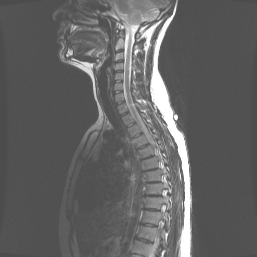
The cervical vertebrae demonstrates hemangioma within the T2 vertebral body with increased T1 and T2 signal and decreased signal on STIR sequence. Bone marrow signal otherwise unremarkable. The cervical cord is normal in caliber and signal. The cranocervical junction is normal.
C3-C4 Right-sided bony ridge and uncinate spurring causing mild right foraminal narrowing. No spinal stenosis
C4-C5 Moderate disc space narrowing. Disc bulge and bony ridge. Posterior ligament hypertrophy. Disc osteophyte complex and posterior ligament hypertrophy causes mild narrowing of the canal and CFS space around the cord. Mild cord flattening. Uncinate spurring with mild bilateral foraminal narrowing.
C5-C6 Small central protrusion. Mild bony ridge. Mild impression ventral subarachnoid space. No spinal stenosis. No significant foraminal narrowing.
C6-C7 Normal. (finally ! something is normal!)
C7-T1 Disc bulge with left-sided protrusion. Mild foraminal narrowing.
Impression: Multilevel spondylosis, as above.
Well... I am no doctor... but this does not sound good. I am a very analytical person, if you could 'decipher' these for me I'll be very grateful.
Thank you,
Carlos
AnswerThe cervical vertebrae demonstrates hemangioma within the T2 vertebral body with increased T1 and T2 signal and decreased signal on STIR sequence. Bone marrow signal otherwise unremarkable. The cervical cord is normal in caliber and signal. The cranocervical junction is normal.(Hemangiomas are benign tumors in the bone that show up as irregularities on x-rays. They are usually nothing to worry about so don't let the word "tumor" cause panic. Everything else looks good.)
C3-C4 Right-sided bony ridge and uncinate spurring causing mild right foraminal narrowing. No spinal stenosis (You've got bone spurs or jagged edges off the bones in your neck that are making the space where the nerves exit tighter.)
C4-C5 Moderate disc space narrowing. Disc bulge and bony ridge. Posterior ligament hypertrophy. Disc osteophyte complex and posterior ligament hypertrophy causes mild narrowing of the canal and CFS space around the cord. Mild cord flattening. Uncinate spurring with mild bilateral foraminal narrowing.(Same as above, but you also have a disc that is pushing backwards along with thickening of the ligaments there, all of which are essentially "choking" the spinal cord and exiting nerve roots. Bottom line is there isn't enough space for the cord and nerves here.)
C5-C6 Small central protrusion. Mild bony ridge. Mild impression ventral subarachnoid space. No spinal stenosis. No significant foraminal narrowing.(The center of the disc "jelly" has leaked out a little, but it isn't putting any pressure on the spinal cord.)
C6-C7 Normal. (finally ! something is normal!)
C7-T1 Disc bulge with left-sided protrusion. Mild foraminal narrowing. (At the very bottom of your neck, you have another disc that has leaked out to the left side making less space for the nerve there.)
Based on these MRIs, I would imagine you are experiencing some pain in your neck as well as some pain and perhaps some numbness going down into your arms or shoulder blades. I think your doctor is going to recommend you undergo some physical therapy to see if it will reduce some of your symptoms, which I believe is the way to go before considering any other more invasive options. Good luck!