Supracondylar femur fractures are becoming more common as the population ages. These fractures usually occur in elderly patients with multiple comorbidities and osteoporotic bone; thus, a high rate of complications exists.[1]
The goal in treating supracondylar femur fractures, as in treating any periarticular fracture in a weightbearing bone, is restoration of a stable limb for functional, pain-free ambulation. Initially, fixation and, finally, healing of the bone restores stability. Maintaining anatomic alignment and length and preventing stiffness restore function. Avoiding arthritis, which requires restoration of anatomic congruent joint surfaces and maintaining the normal mechanical axis of the limb, prevents pain.
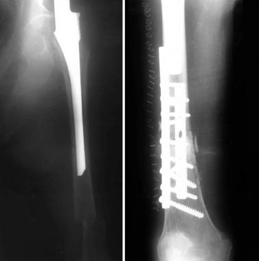
Supracondylar femur fractures require anatomically stable internal fixation for best results. Historically, traction achieved adequate results for the treatment of these fractures; however, the outcomes probably would not be considered acceptable today. Maintaining leg length and preventing varus malalignment is difficult with traction. Although surgical risks were avoided, the patient was exposed to the risks of prolonged bedrest, including pulmonary complications, deep venous thrombosis, pressure ulcers, disuse osteoporosis, and generalized muscle atrophy and deconditioning.[1]
All current authors agree that the best results are now achieved with operative methods.[2] Involvement of the articular surface demands a congruent anatomic reduction to prevent or minimize posttraumatic arthritis and provide bone stock for later knee replacement or fusion.[3, 4]
Severe comminution often requires fixation of multiple independent fragments with one device to minimize soft-tissue damage.[5] Severely comminuted distal femur fractures are especially hard to treat properly.[5, 6, 3, 7, 1, 4, 2, 8, 9] Obtaining adequate fixation may be technically challenging, especially when multiple fragments are present. The significant forces applied to this area, even during restricted patient activities, require a strong implant; however, fixation is difficult because of the wide canal, the thin cortex, and the relatively poor bone quality of the distal femur.[7, 8, 10]
Most surgical failures are caused by inadequate fixation of fracture fragments.[11] Each device has limitations, and no implant can stabilize every fracture type; however, for best results, the device chosen must provide fixation rigid enough for early motion.[5, 4, 12, 13] If comminution and the fracture pattern compromise the use of an implant, the surgeon should be flexible and choose the device that fits best.[14]
Supracondylar femur fractures that occur after total knee replacement are also more difficult to treat adequately because the knee replacement prosthesis can interfere with fixation implants.[15, 16, 17]
NextThe distal femur is funnel-shaped, and the area where the stronger diaphyseal bone meets the thinner and weaker metaphyseal bone is prone to fracture with direct or indirect trauma. The surgeon needs to be aware of the shape of the bone when planning surgery so that the implant matches the bone.
The approach to the thigh is a standard lateral one, with an incision through the fascia lata and access to the bone along the intermuscular septum under the vastus lateralis. The femoral artery is medial; other neurovascular structures are posterior and thus should not be encountered during surgery.
Supracondylar femur fractures usually occur as a result of low-energy trauma in osteoporotic bone in elderly persons or of high-energy trauma in young patients. Fractures proximal to knee replacements may be caused by notching of the anterior cortex when the surgeon placed the prosthesis or may be secondary to the stress riser effect of the interface between the rigid metal and soft bone.[18] The treating physician must also be aware of the potential for pathologic fractures through metastatic lesions or primary bone tumors in this area.
With stable fixation, anatomic alignment, and restoration of intra-articular congruency, most patients do well. The more comminuted the fracture and the poorer the quality of bone, fixation, or reduction, the worse the prognosis. Severe comminuted type C3 fractures are expected to develop significant stiffness and posttraumatic arthritis. Patients with open fractures fare worse than those with closed fractures.
Periprosthetic fractures and dementia, heart failure, advanced renal disease, and metastasis lead to reduced survival. Dealying surgery for longer than 4 days leads to increases in 6-month and 1-year mortality. Mortality after native fractures of the distal femur in the geriatric population is high and is comparable to mortality after hip fractures.[19]
Clinical Presentation
Copyright © www.orthopaedics.win Bone Health All Rights Reserved