Giant cell tumors of the tendon sheath are the second most common tumors of the hand, with simple ganglion cysts being the most common.[1] Chassaignac first described these benign soft-tissue masses in 1852, and he overstated their biologic potential in referring to them as cancers of the tendon sheath.
Giant cell tumors of the soft tissue are classified into the following two types:
The rare diffuse form is considered the soft-tissue counterpart of diffuse pigmented villonodular synovitis (PVNS) and typically affects the lower extremities.[2] Its anatomic distribution parallels that of PVNS, with lesions most commonly found around the knee, followed by the ankle and foot; however, it occasionally affects the hand. Typically, these lesions, like those of PVNS, occur in young patients; 50% of cases are diagnosed in patients younger than 40 years. The diffuse form is often locally aggressive, and multiple recurrences after excision are common.
Because of the similarities in age, tumor locations, clinical presentations, and symptoms for patients with PVNS and patients with the diffuse form of giant cell tumors of the tendon sheath, the diffuse form probably represents an extra-articular extension of a primary intra-articular PVNS process.
Findings from flow cytometric DNA analysis suggest that PVNS and giant cell tumors of the tendon sheath are histopathologically similar but clinically distinct lesions.[3, 4, 4] When the origin of these poorly confined soft-tissue masses is uncertain, Enzinger and Weiss[5] classify these tumors as the diffuse type of giant cell tumors of the tendon sheath, whether or not they involve the adjacent joint.[6]
This article focuses on the common localized form of giant cell tumors—that is, the giant cell tumors of the tendon sheath that are often found in the hands and feet.[7, 8, 9, 10, 11]
NextAs is true for most soft-tissue tumors, the etiology of giant cell tumors of the tendon sheath is unknown. Pathogenetic theories have included trauma, disturbed lipid metabolism, osteoclastic proliferation, infection, vascular disturbances, immune mechanisms, inflammation, neoplasia, and metabolic disturbances.[12] Probably the most widely accepted theory, as Jaffe et al proposed,[13] is that of a reactive or regenerative hyperplasia associated with an inflammatory process.
Histochemical evidence shows that the mononuclear cells and giant cells present in these lesions resemble osteoclasts,[14, 15] and this resemblance suggests a bone marrow–derived monocyte/macrophage lineage for these tumors. Polymerase chain reaction (PCR) assays have shown that giant cell tumors of the tendon sheath are polyclonal proliferations,[16] which suggests that these masses are nonneoplastic proliferations, if one accepts the premise that a population of cells forming a tumorous mass must show clonality to be classified as a neoplasm.
Giant cell tumors of the tendon sheath are the second most common tumors in the hand; simple ganglion cysts are the most common. Giant cell tumors of the tendon sheath most commonly occur in patients aged 30-50 years, with a peak incidence in those aged 40-50 years. Rarely are these tumors found in patients younger than 10 years or older than 60 years. The female-to-male ratio is 3:2.
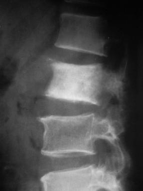
Giant cell tumors of the tendon sheath are associated with degenerative joint disease, especially in the distal interphalangeal (DIP) joint. Jones et al[17] noted degenerative joint disease in the joint from which a tumor arose or in the joint nearest to the mass in 46 of 91 cases in which radiographs were reviewed.
An occasional association with rheumatoid arthritis has been reported;[18] however, to the authors' knowledge, no pathogenetic relation between rheumatoid arthritis and giant cell tumor of the tendon sheath has been demonstrated, and their simultaneous occurrence may be coincidental. Antecedent trauma occurs in a variable number of these patients, but its association with these tumors is also probably coincidental.
The incidence of local recurrence is high, ranging from 9% to 44%. Researchers have reported the following rates:
The variability in rates probably reflects incomplete excision of the lesions, especially the satellite nodules. Risk factors for recurrence include the following:
Goda et al have presented a new technique for the use of radiotherapy as an adjuvant modality to prevent local recurrence.[23] For retrospective studies, see Rodrigues et al,[24] Darwish and Haddad,[25] and Messoudi et al.[26] For a significant study in children, see Gholve et al.[27]
To the authors' knowledge, no cases of malignant degeneration of a benign giant cell tumor of the tendon sheath of the hand have been reported. These tumors also have no propensity to metastasize distally. A few sporadic cases of purported malignant giant cell tumors have been reported; however, most authors doubt that these malignant tumors exist, because this diagnosis is difficult to confirm.
Clinical Presentation
Copyright © www.orthopaedics.win Bone Health All Rights Reserved