The aneurysmal bone cyst (ABC; see the image below) is an expansile cystic lesion that most often affects individuals during their second decade of life and may occur in any bone in the body.[1, 2, 3, 4, 5, 6, 7, 8, 9, 10, 11] Although benign, the ABC can be locally aggressive and can cause extensive weakening of the bony structure and impinge on the surrounding tissues.
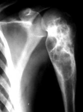 Aneurysmal bone cyst of the upper arm. Courtesy of Johannes Stahl, The Virtual Radiological Case Collection.
The true etiology and pathophysiology remain a mystery, but the mainstay of treatment has been intralesional curettage.[12] Recurrence is not uncommon.[1, 13] Other surgical options include en-bloc resection or wide excision, selective arterial embolization, and curettage with locally applied adjuvants such as liquid nitrogen, argon beam photocoagulation, or phenol.
NextJaffe and Lichtenstein first described ABC as its own entity in 1942, when they noted "a peculiar blood-containing cyst of large size."[14] Two cases were reported in which a lesion with a "soap-bubble" appearance on radiographs was found on the superior pubic ramus of a 17-year-old male and on the second vertebrae of an 18-year-old male. The lesions were expansile and showed evidence of erosion of the surrounding bone and encroachment of the surrounding tissues. Upon surgical exposure of the lesions, a thin, bony wall that contained bloody fluid was found.
Jaffe and Lichtenstein suggested that ABCs may have been mistaken for other benign and malignant bone tumors in the past.[14] Although ABC is a separate entity, in some situations, distinguishing an ABC from a giant cell tumor of bone or a telangiectatic osteosarcoma is difficult.
As defined by the World Health Organization, the ABC is a benign tumorlike lesion.[4] It is described as "an expanding osteolytic lesion consisting of blood-filled spaces of variable size separated by connective tissue septa containing trabeculae or osteoid tissue and osteoclast giant cells."[4] Although benign, an ABC can be a rapidly growing and destructive bone lesion. Its expansile nature can cause pain, swelling, deformity, disruption of growth plates, neurologic symptoms (depending on location), and pathologic fracture.[1, 2, 3]
ABCs are generally considered rare, accounting for only 1-6% of all primary bony tumors. A group from Austria reported an annual incidence of 0.14 ABCs per 100,000 people[15] ; however, the true incidence is difficult to calculate because of the existence of spontaneous regression and clinically silent cases.
A biopsy-proven incidence study from the Netherlands showed that ABCs were the second most common tumor or tumorlike lesion found in children.[16]
Most studies have also found a slightly increased incidence in women. Although the ABC can appear in persons of any age, it is generally a disease of the young (albeit a rare one in the very young). About 50-70% of ABCs occur in the second decade of life, with 70-86% occurring in patients younger than 20 years. The mean patient age at onset is 13-17.7 years.
The true etiology of ABCs is unknown. Most investigators believe that ABCs are the result of a vascular malformation within the bone; however, the ultimate cause of the malformation is a topic of controversy. Three commonly proposed theories are as follows:
A certain percentage of primary ABCs may be truly neoplastic — as opposed to vascular, developmental, or reactive — phenomena. It has been shown that as many as 69% of primary ABCs demonstrate a characteristic clonal t(16;17) genetic translocation[17] leading to upregulation of the TRE17/USP6 oncogene,[18] whereas no secondary ABCs demonstrate this cytogenetic aberration.
The true pathophysiology of ABCs is also unknown.[12]
Different theories about several vascular malformations exist; these include arteriovenous fistulas and venous blockage. The vascular lesions then cause increased pressure, expansion, erosion, and resorption of the surrounding bone. The malformation is also believed to cause local hemorrhage that initiates the formation of reactive osteolytic tissue. Findings from a study in which manometric pressures within the ABCs were measured support the theory of altered hemodynamics.
Most primary ABCs demonstrate a t(16;17)(q22;p13) fusion of the TRE17/CDH11-USP6 oncogene. This fusion leads to increased cellular cadherin-11 activity that seems to arrest osteoblastic maturation in a more primitive state.[18] This process may be the neoplastic driving force behind primary ABCs as opposed to secondary ABCs, that seem to occur reactively as a result of another underlying disease process.
Patients usually present with pain, a mass, swelling, a pathologic fracture, or a combination of these symptoms in the affected area. The symptoms are usually present for several weeks to months before the diagnosis is made, and the patient may also have a history of a rapidly enlarging mass. Neurologic symptoms associated with ABCs may develop secondary to pressure or tenting of the nerve over the lesion, typically in the spine.
Pathologic fracture occurs in about 8% of ABCs, but the occurrence rate may be as high as 21% in ABCs that have spinal involvement.
Other findings may include the following:
ABCs are generally treated with surgery. Rarely, asymptomatic ABCs may be seen in which there is clinically insignificant destruction of bone. In such cases, close monitoring alone of the lesion may be indicated because of the evidence that some ABCs spontaneously resolve. When a patient is monitored in this manner, the diagnosis must be certain, and the lesion should not be increasing in size.
Some anatomic locations may be difficult to access surgically. If this situation is encountered, other methods of treatment, such as intralesional injection and selective arterial occlusion, may be successful.
Impending pathologic fracture, especially a fracture of the hip, is a challenging problem and an indication for intervention, which often includes curettage, adjuvant treatment, and internal fixation.
ABCs may affect any bone in the body; thus, the relevant surgical anatomy varies with location. ABCs most commonly affect the long, tubular bones, followed by the spine and flat bones. These three areas account for 80% of all ABCs. When present in long, tubular bones, ABCs tend to be eccentrically located in the metaphysis.
ABCs least commonly involve a subperiosteal location, where they may form a predominant soft-tissue mass. However, ABCs can occur in any location, including the diaphysis and epiphysis.
Rarely, ABCs have also been known to affect an adjacent bone; however, spinal ABCs are associated with a higher incidence of contiguous lesions. Almost all ABCs of the spine involve the posterior elements, and a high incidence of neurologic symptoms is observed, as well as more local aggressive behavior.
The pelvis accounts for approximately 50% of lesions occurring in the flat bones.[19] Secondary lesions tend to have a predilection for the areas of the body in which the primary lesion typically arises.
In a published review of 897 cases of ABC, the following rates of occurrence were reported[20] :
Contraindications for selective arterial embolization include the following:
Contraindications for intralesional injection are as follows:
Contraindications for radiotherapy include the following:
Concerns for local resection include the following:
Concerns for en-bloc excision of a deep lesion include the following:
Concerns for intralesional removal include the following:
Concerns for adjuvant intralesional therapy include the following:
Copyright © www.orthopaedics.win Bone Health All Rights Reserved