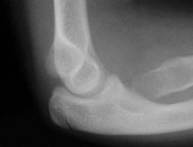
Pilon is a French word for pestle, an instrument used for crushing or pounding.[1] The first recorded use of the term pilon in the orthopedic literature is in 1911, by Étienne Destot.[2] Pilon fractures account for approximately 7% of tibial fractures. Pilon fractures in the distal tibia result from axial forces that can range from low to high energy and produce a spectrum of articular and metaphyseal injuries. These fractures involve the dome of the distal tibial articular surface and extend into the adjacent metaphysis. The fibula may or may not be intact.
Despite the advances that have been made in management, these fractures can be challenging to deal with, especially when associated with significant soft-tissue injury. Although various options are available to treat these fractures, timing of definitive surgery is crucial with respect to the condition of the soft tissues. New developments in the field continue to lead to better outcomes.
The treatment of pilon fractures has evolved over the last century. Conservative management gave way to surgical intervention when implants became available, but poor outcomes led to a return to cast immobilization or limited internal fixation of the fibula only. However, outcomes after nonoperative treatment continue to be poor.
In 1959, Jergesen stated that open reduction and stabilization of severe tibial pilon fractures is impossible. In the early 1960s, the Association for Osteosynthesis/Orthopaedic Trauma Association (AO/OTA) developed general guidelines for the treatment of intra-articular distal tibial fractures, which led to open reduction and anatomic and rigid internal fixation.
Good outcomes were reported when these principles were used for low-energy injuries (eg, those from skiing accidents). However, when the same principles were used to fix high-energy injuries (eg, from motor vehicle accidents), the outcomes were poor, mainly because of soft-tissue complications.
Over the years, the importance of soft tissues and the differences between low- and high-impact energy injuries have become better understood. This has led to the development of newer treatment concepts, which continue to evolve, along with an availability of more advanced surgical options, such as minimally invasive internal fixation implants.[3, 4, 5]
NextThe distal tibia and fibula, along with the ligaments and capsule, help to form the ankle mortise. Any disruption of length, axis, or rotation of the fibula or the tibia can result in an incongruent ankle joint.
The lateral aspect of the distal tibia forms a triangular notch, which is where the fibula articulates. The interosseous and the anterior and posterior tibiofibular ligaments bind these bones together.
The ligaments often avulse fragments from the tibia, such as the anterolateral fragment termed the Chaput fragment and the posterior malleolar fragment termed the Wagstaffe fragment.[6]
The blood supply in the distal leg is provided by branches that arise from the posterior tibial, peroneal, and dorsalis pedis arteries.
The great saphenous vein travels along with the saphenous nerve anterior to the medial malleolus. The small saphenous vein passes posterior to the lateral malleolus. Disruption of the venous system can lead to subsequent chronic venous stasis.
Depending on the mechanism of injury and the direction of forces, a wide variety of injury patterns result. At one end of the spectrum are low-energy injuries that follow activities such as skiing and result in minimal soft-tissue injury. The fracture fragments are fewer, have a spiral orientation, and are relatively minimally displaced.
At the other end of the spectrum are high-energy injuries such as a fall from height or a high-speed motor vehicle accident. Such a mechanism can produce significant comminution with multiple displaced fracture fragments and, importantly, a contused or crushed soft-tissue envelope, which could also be breached and open to external contamination through wounds. The fibula is usually fractured in high-energy injuries.
A variable amount of damage can occur to the articular cartilage of the tibia, which can be scuffed, bruised, or fragmented. In severe cases, the weightbearing central dome can be fragmented. The fragments, which can be tiny (~2-3 mm3), are completely broken off and are driven up into the metaphysis of the tibia by the impact. Damage to the talar articular surface can also occur.
Pilon fractures occur when the talus is driven into the tibial plafond. The forces that cause fracture fragments in the distal tibial metaphysis, articular and soft-tissue damage, can be vertical, rotational, angular, or a combination of these.
The outcome varies, depending on the following factors:
Low-impact pilon fractures have better outcomes than high-impact pilon fractures. In general, good outcomes can be expected in approximately 60-80% of patients.[7, 8, 9, 10, 11]
Many patients continue to improve for many years after the injury. The severity of the injury and the quality of the articular reduction frequently correlate with the development of arthrosis, but radiographic signs of arthrosis have only a weak correlation with clinical outcome[12] .
Ankle fusions may be required in approximately 3-27% of patients with posttraumatic arthritis. Nonunion in the distal tibia can be treated with a fibula-pro-tibia plating and bone grafting procedure, as described by DeOrio and Ware.[13] Ankle replacement is an option in selected individuals.
Outcomes following an innovative approach using staged posterior tibial plating for the treatment of OTA 43C2 and 43C3 pilon fractures in 9 patients with a separate, displaced, posterior malleolar fragment were compared by Ketz and Sanders to 10 patients with similar fracture patterns treated using standard anterior or anteromedial incisions with indirect reduction of the posterior fragment.[14] All 19 patients were available for follow-up at an average of 40 months (range, 28-54 months). The average Maryland Foot Score and American Orthopaedic Foot and Ankle Society (AOFAS) Ankle and Hindfoot score for the first group were 86.4 and 85.2, compared with 69.4 and 76.4 for the later group.
The authors concluded that the addition of a posterior lateral approach offers direct visualization for reduction of the posterior distal fragment of the tibial pilon.[14] Although the joint surface itself cannot be visualized, this reduction allows the anterior components to be secured to a stable posterior fragmentat a later date. This technique improved ability to subsequently obtain an anatomic articular reduction based on computed tomography (CT) scans and preservation of the tibiotalar joint space at a minimum 1-year follow-up.[14]
Vidović et al reported on a prospective study involving 21 patients with closed distal tibial and pilon fractures treated with minimally invasive plate osteosynthesis (MIPO).[15] Nineteen patients were initially managed with an ankle-spanning external fixator. When the status of the soft tissue had improved and swelling had subsided enough, a definitive internal fixation with MIPO was performed. Patients were invited for follow-up examinations at 3 and 6 weeks and then at intervals of 6 to 8 weeks until 12 months. Mean age of the patients was 40.1 years (range 19-67 years). Eighteen cases were the result of high-energy trauma and three were the result of low-energy trauma.
By the AO/OTA classification, there were extra-articular and intra-articular fractures, but only simple articular patterns without depression or comminution.[15] The average time for fracture union was 19.7 weeks (range, 12-38 weeks). Mean range of motion was 10° of dorsiflexion (range, 5º-15°) and 28.3° of plantar flexion (range, 20º-35°). Three cases were metalwork-related complications. Two patients underwent plate removal at 24 weeks because of plate impingement. There was one case of wound breakdown at 11 weeks. One patient had fracture union with tibial recurvatum of approximately 10°, without functional impairment. Two patients had delayed union.
Vidović et al concluded that MIPO is a reliable method of treatment for distal tibial fractures; it provides a high union rate and good functional outcome with minimal soft-tissue complications.[15] Skin impingement remains a common complication with MIPO, but this can be solved by timely plate removal.
Clinical Presentation
Copyright © www.orthopaedics.win Bone Health All Rights Reserved