QuestionHonorable Doctor
I am General Practitioner (GP) in Pakistan; one of my friends is really in a great trouble and facing the annoyance of spine surgery which couldn't be succeeded due to unknown reasons, so that he is in need of your kind help & competent advice purely on compassionate grounds, having your vast experience/knowledge in the field of Neuro surgery. Please furnish the answers of Queries given below in the light of following history, for which I shall ever pray for your long life and prosperity.
PRE-OPERATIVE HISTORY
He is 46 years old male and suffering from Low back pain along with left sciatica since September 2001, while the pain was constant in nature and radiated towards buttock/left leg and became worst on sitting, standing and getting out of bed, as such there was no neurological deficit. The SLR of Rt leg & Lt leg was 70 &50 degrees respectively. The conservative treatment including NSAID's and physiotherapy received but got no relief.
POST-OPERATIVE HISTORY
Due to failure of conservative treatment and having more than three years of back pain history he ultimately entered into lumbar surgery in February 2005 on the advice of concerned Neuro surgeon, because the result of his MRI showed multilevel disc degeneration & at the Level of L4-5 there was mild central posterior disc protrusion causing pressure on ventral surface of thecal sac. The surgeon used the technique of Fenestration and removed the disc at L4-5 without doing a fusion, but unfortunately the LEG/LOW BACK PAIN couldn't be subsided till now & intensity of pain is same as pre-op, beside using NSAID's/muscle relaxant/anti seizure medicines/Facet joint injection & having postoperative physiotherapy. Now the pain is worsened by sitting, standing or walking for a long period of time. Continued Left leg stiffness and back discomfort also persists & badly distressing his routine life.
However he repeated his MRI scan with contrast along with X-rays of L/spine after operation and the reports of the same are reproducing below for your kind perusal.
MRI LUMBOSACRAL SPINE DONE WITHOUT AND AFTER CONTRAST ENHANCEMENT WITH GADOLINIUML:
Spin Echo T1 weighted and gradient rephased T2 weighted images were obtained in axial and sagittal plane.T1 weighted images were obtained after contrast enhancement in axial, sagittal and coronal planes.
HISTORY:- History of surgery on 11th feb.2005 at the level of L4/5.Films were compared with previous scan of 11th Feb.2005.
*There is disc degeneration in the lumbar spine at L2/3 and L4/5 with narrowing of disc space at L4/5.
* Previously noted diffuse disc herniation at L4/5 appears resected.Now there is disc bulging with facet joint hypertrophy at L4/5 causing slight pressure over thecal sac and left neural foramina.
* Postoperative changes are noted at the level of L4/5 in the posterior paraspinal region along with enhancing granulation tissue within the thecal sac and posteriorly at this level.
* Disc bulging is noted at L3/4 and L5/S1.
* The spinal cord shows no abnormal signal.
* The vertebral bodies show no evidence of abnormal signal to indicate bone marrow replacement.
* There is no spinal stenosis.
* MR myelogram shows no obstruction to flow of CSF.
CONCLUSION:-
* Previously noted diffuse disc herniation at L4/5 appears resected.Now there is disc bulging at this level with facet joint hypertrophy causing slight narrowing of left neural foramina.
*There is also disc bulging at L3/4 and L5/S1.
*There is no spinal stenosis.
* MR myelogram shows no obstruction to flow of CSF.
LUMBOSACRAL SPINE (AP & LAT):--
* Loss of normal lumbar lordosis.
* Osteophytes are seen along the margins of vertebral bodies.
* Partial sacralization of L5 vertebra is seen.
* Inter-vertebral disc spaces appear normal.
* No fracture or dislocation is seen.
* No bony erosion is seen.
* Paravertebral soft tissue shadows appear normal.
* S.I joints appear normal.
IMPRESSION:-
* Loss of normal lumbar lordosis梔ue to muscle spasm.
* Osteoarthritic changes seen in lumbar spine.
* Partial sacralization of L5 vertebra.
QUERIES: ---
1. The report of post-op MRI shows that there is disc bulging at L4-5 level with facet joint hypertrophy causing slight narrowing of left neural foramina. How this narrowing occurred abruptly after surgery, as this was not identified before operation? Is it usually happens in this kind of operation or it is an error at the part of concerned Neuro surgeon?
2. What probable cause of persistent Leg/LBP can be assessed from post-op MRI/X-rays report?
3. Is it seems to be inadequate surgery OR wrongly selection of the patient?
4. What treatment/advice does u suggest at this stage to alleviate this constant pain without intervention of repeat surgery? AND if repeat surgery is the only option than how much risk could be involved in it & how much success can be expected apparently?
5. The concerned surgeon again inducing him to enter into second surgery on the basis of following version: ----
?Unfortunately the intervertebral canal from where nerve passes was little longer in your case as routine practice. I cleared it medially but the lateral part shows little obstruction to nerve. This is not failed back surgery that an other phenomenon but obstruction to nerve in lateral part of foramen, some times residual disc pain, Discitis and lateral discs problems are well established and well acknowledged in the world where repeated surgery is advised. I advice you to have EMG done so that I pinpoint that nerve and then request you for minor procedure again to open the nerve canal. I will just do 1/2 hour procedure to clear your nerve root more distally and relive your pain. ?br>
6. Whether the above narration seems to be believable? In this connection your valuable comments would certainly help to redress the frustration of my distressed friend.
Looking forward to your expertise?br>
Thanks,
GUL KHAN
AnswerDr. Gul Khan:
Thanks for writing!
Indeed, an interesting case.
To answer your question. Question # 1; The narrowing is a side effect of the surgery. The surgeon did nothing wrong, they followed current procedure.
#2: The cause of the persistent low back and leg pain is the postion of the bones and disc and ligaments of the low back. They are postioned so that the keep the dura mater from moving as it should.
#3: It is a situation where the condition of the patient did not meet the requirements for a successful back surgery.
The only time back surgery is successful is when the patient's spinal cord is contracted, that is, the top end is closer to the bottom end than it used to be. This is very often apparent in loss of height in the patient. See if you can find in the charts a measurment of the patient's height just before the back surgery. Compare that to the height the patient reports 10 years before the back surgery.
If the height of the patient just before the back surgery is less than the previous height, the back surgery should have been successful. I would wager that he did not loose height.
#4 I suggest at this time doing the following. Increase the amound of water intake of the patient. It has to reach a minimum level. For example, if he weight 220 lbs, he must take in 110 ounces of water a day. Other liquids do not cound, just filtered water.
The get this book. "How to deal with back pain and Rheumatoid Joint Pain" by Batmanghelidj. There are postioning procedures in there which are very effective in dealing with a situation such as this.
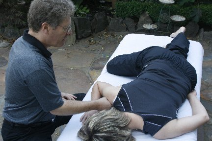
In addition, have the patient do this exercise. Lie on their back, keep the left leg straight, move it out away from their body as far as they can (abduction) and then rotate the toe so that it points away from them. This will very likely make the pain much worse the first time. Rest for two minutes, they try it again. If the pain get better, the problem was a piriformis syndrome and the problem is now solved. It can be redone as neccessary to control pain.
Do not repeat this manuever if it hurts every time the patient does it. Just try it for three or four applications. If the pain get worse each time, it is not the thing to do.
Of course the diet is important. Get the patient to eat as much chicken bone gristle as possible, or other animal gristle, if they are not vegetarian. The active ingredient is chrondroition glucoamine sulfate, perhaps it is available in pill form, but the direct source is better.
Does this answer your question?
I see that your patient also posted a question to me but I will not answer that, since I have covered everything here, it think.
Dr. Rozeboom