Toe walking is a gait abnormality characterized by an absence of normal heel-to-floor contact (heel strike) by both feet during gait, with the forefoot engaging in the majority of floor contact throughout the gait cycle. Toe walking has multiple etiologies, ranging from idiosyncratic habit to profound neuromuscular disease. Critical to the management of toe walking is the exclusion of neurologic or muscular diseases as a cause of the perceived gait abnormality. Treatment depends on the patient’s age and severity of the gait abnormality. Specific treatment options range from simple observation to surgical lengthening of muscles or tendons in the lower extremity.
NextTenotomies are the commonly used procedure for the treatment of toe walking. It is also one of the oldest procedures in orthopedics. The first tenotomy is believed to be a multilevel percutaneous Achilles tendon lengthening performed by Delpech in 1823 while treating a patient with clubfoot.[1]
Toe walking is defined as the inability to make heel contact with the floor (heel strike) during the initial stance phase of the gait cycle and the absence of full foot contact with the ground during the remainder of the gait cycle. Although toe walking is commonly seen during development in children who are first learning to walk, a consistent heel-toe pattern of gait usually develops by approximately age 22 months.[2, 3] Persistent toe walking beyond age 2 years merits further evaluation.[4]
The most commonly observed type of toe walking is idiopathic toe walking (ITW). The true prevalence of ITW is unknown because not all children with ITW present to the doctor. In several small studies, ITW has been estimated to occur in 7-24% of the childhood population.[5] A large cross-sectional study from 2011 from the Netherlands found a prevalence of 12% in the general population.[5] Another large study from 2012 in Sweden found the prevalence of ITW to be 4.9% in children aged 5 years 6 months.[6]
ITW is observed with a higher frequency in patients with autism, developmental delay, and language disorders.[7] A recent study determined the incidence of toe walking in children with autistic spectrum disorder to be 20.1%, while historical reports estimate it to be as high as 63%.[8] Conversely, children with ITW display an increased prevalence for other pediatric neuropsychiatric disorders such as attention-deficit/hyperactivity disorder, tics, Tourette syndrome, and obsessive compulsive disorder.[9]
The most common identifiable etiology of toe walking is cerebral palsy, which affects 1-7 in 1000 children. Reports vary greatly regarding the incidence of toe walking in this population, as disease severity spans a spectrum. However, it is noted that less than 50% of these patients overall walk on their toes.[10]
Duchenne muscular dystrophy has an incidence of 1 case per 3500 live male births and is the most common degenerative muscle disease of childhood.[11] The typical child with Duchenne muscular dystrophy initially walks with a heel-to-toe pattern but progresses to a consistent toe-walking pattern as a compensation for the progressive weakness of the knee extensors.
Various causes of toe walking exist and include both central and peripheral neurologic disorders (eg, tethered cord, diastematomyelia, spina bifida, cerebral palsy), muscular disorders (eg, congenital muscular dystrophy), and anatomic disorders such as limb length discrepancy.[4] Some rarer causes of toe walking have also been reported in the literature, such as acute toe walking secondary to viral myositis in a previously healthy 4-year-old child.[12]
The most common cause of toe walking is idiopathic, meaning no identifiable pathologic process exists to explain the perceived gait abnormality. Idiopathic toe walking (ITW), first described by Hall in 1967 as “habitual toe walking” and “congenital short Achilles tendon,” is a diagnosis of exclusion.[13] ITW is best defined as bilateral toe walking with or without Achilles tendon contracture in a child older than age 2 years in the absence of other etiologies.
Physical Medicine and Rehabilitation for Spasticity
Spinal Cord Trauma and Related Diseases
Muscular Dystrophy
Congenital Myopathies
Although the exact pathophysiology of idiopathic toe walking (ITW) remains unknown, it is postulated that mild defects in sensory processing exist in affected children and that this gait may result from a vestibular disorder or abnormal sensitivity to touch.[14] However, there is limited research exploring these relationships. ITW likely has a genetic component since a positive family history has been reported in many case series.[7] In the initial description of the condition, Hall et al noted that all their patients had congenital shortness of the Achilles tendon, which led to ankle equinus and toe walking.[13] However, subsequent studies have demonstrated that not all patients with ITW exhibit a congenitally short Achilles tendon as a mechanical explanation for the gait difference and some patients toe walk despite a volitional ability to walk flat-footed (“dynamic toe walkers” or “habitual toe walkers”).[7]
The natural history of ITW remains poorly defined as most studies offering long-term follow-up concurrently report treatment of their cohorts. Opinion on the adult consequences of a persistent toe-walking pattern may be divided into two schools of thought.
In the first, it is believed that regardless of the initial status of the heel cord and ankle range of motion, children with persistent toe walking eventually develop a fixed ankle contracture and ankle equinus in adulthood, leading to hindfoot valgus and myriad potential foot disorders.[15, 16, 17, 18, 19] This argument supports aggressive intervention for ITW in early childhood. In the second school of thought, it is believed that ITW can have a benign natural history, with the majority of patients manifesting no particular functional limitations or pathologic sequelae as an adult, despite variable objective improvement in overall ankle motion or gait.[20] The second viewpoint supports an observational approach to management of this gait abnormality.
The pathophysiology of toe walking in patients with cerebral palsy is clearer and relies on two basic mechanisms centered on the underlying spasticity of the lower extremity musculature. First, spasticity of the foot and ankle muscles can lead to progressive ankle equinus contracture as spastic muscles grow at a slower rate than muscles that are not spastic.[21] Second, spasticity and flexion of the more proximal hip and knee joints can result in apparent toe walking as the patient attempts to maintain balance when upright. If the hip and knee are flexed in stance and the ankle is held at a right angle relative to the tibia (plantigrade), the patient bears weight on the toes and forefoot, even though the ankle itself is not in equinus.[21]
In Duchenne muscular dystrophy, the quintessential example of degenerative muscle disease, lower extremity muscles progressively weaken as they degenerate and are replaced by fibrous tissue. Toe walking results from the relatively greater involvement of the dorsiflexors rather than the plantar flexors of the foot. In addition, as noted previously, toe walking also develops to compensate for the weakening quadriceps muscle. As the quadriceps becomes weak, active knee extension is lost and knee stability during gait is threatened as the knee preferentially buckles into flexion. By walking on the forefoot, the patient generates a knee extension moment that aids in its stability.[22]
Children with idiopathic toe walking (ITW) typically present as a toddler, without other significant medical history and with a normal developmental profile, especially a normal age for the commencement of walking (before 18 months). The parents report that the child walks and runs on the toes of both lower extremities symmetrically, particularly when he or she is unaware of being observed. However, the child often can walk flat-footed if prompted.
The presentation of toe walking in patients with discrete central or peripheral nervous system etiologies differs from that of the patient with ITW. In patients with cerebral palsy, there is often a history of prematurity and of global developmental delay, especially a delayed age at which the child began walking. There may also be a history of a significant head injury or vascular event before age 2 years. Other features upon presentation that may suggest a neurologic etiology for toe walking include a history of spinal cord injury, progressive loss in lower extremity function, or asymmetric progressive lower extremity deformity. Specific spinal cord lesions that can result in asymmetric toe walking include spinal cord tumor or split-cord malformation/diastematomyelia.
A patient with toe walking secondary to a degenerative muscle disease usually presents later, from age 3-5 years, often after the primary diagnosis has been established. Developmental history or past medical history in this patient population is typically normal.
The differences in presentation between the various patient groups can be subtle and subject to overlap. Therefore, it is imperative that the examiner routinely reviews the child’s perinatal history, developmental history, and past medical history, as well as obtains a detailed history of present illness, to minimize the likelihood of missing a diagnosis with significant long-term medical implications for the patient.
The examiner’s responsibility in evaluating a patient with toe walking is to rule out all defined etiologies of toe walking before settling on an “idiopathic” diagnosis. The examination should begin with an overall assessment of patient appearance and gait. Gait can be observed as the patient moves about the room or as the patient walks down a hallway. A focused examination of the spine and lower extremities is then required. The child’s lower extremities and spine should be inspected for cutaneous abnormalities, leg-length discrepancy, asymmetric or abnormal muscle development, pelvic asymmetry, and fixed foot deformities.
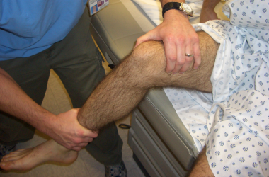
Although a thorough neurologic examination is challenging in this age group, muscle strength can be gauged by the ability of the child to rise independently from the floor and to climb the examination table. Reflexes, muscle tone, and withdrawal to stimuli are easier data points to acquire. Range of motion of the knee, hip, and ankle joints completes the examination. Ankle range of motion should be assessed with both the knee flexed and knee extended (see Silfverskiöld Test in Relevant Anatomy). In addition, ankle dorsiflexion should be examined with the heel in neutral position, as hindfoot valgus gives the false appearance of acceptable dorsiflexion.
For the patient with ITW, the examination is normal except for the symmetric presence of bilateral toe walking and a variable degree of heel cord tightness. An asymmetric toe-walking profile essentially excludes the idiopathic diagnosis and prompts further investigation.
In a patient with a central nervous system disorder, such as cerebral palsy, muscle spasticity is apparent at the time of the initial evaluation and deep tendon reflexes are hyperactive. Gait evaluation suggests overall difficulty with coordination and balance, as well as different degrees of upper extremity flexion posturing, dependent upon the extent of involvement. The examiner should make note of the anatomic distribution of the spasticity, as this is correlated with the extent of the lesion of the motor cortex. In addition, the examiner should determine if the toe walking is secondary to Achilles tendon contracture or as a compensation for spastic knee and hip flexion.
In a patient with muscular dystrophy, the classic findings are lumbar lordosis, calf hypertrophy, and a positive Gower sign (ie, need to prop self up on thighs with arms to rise from a seated position on the floor). Strength examination suggests greater weakness of proximal hip and shoulder muscles.
The management of toe walking is controversial, and there are limited data directly comparing different treatment modalities. Therefore, the physician’s first decision point is whether the toe-walking gait should be treated or if simple observation should be recommended.
Observation is appropriate for a toddler with idiopathic toe walking (ITW) who has recently begun to walk and is without fixed contractures. For many children, this condition represents a temporary habit and they eventually develop a normal heel-toe gait.[2, 3] The patient should be monitored at 6-month intervals. If progressive heel-cord contractures are detected or if the pattern does not resolve spontaneously by age 3 years, treatment can be considered.
If treatment is offered, nonoperative management is always considered before operative management. Nonoperative modalities include stretching, casting, orthotics, and chemodenervation with botulinum toxin. If these are not successful and surgery is chosen, surgical options range from simple heel cord tenotomy or gastrocnemius fascia lengthening to multiple muscle lengthening within the lower extremity. The final surgical approach depends heavily on the underlying pathology of the toe walking.
The triceps surae muscle-tendon complex is the confluence of the gastrocnemius and soleus muscles and the Achilles tendon or heel cord, the largest tendon in the human body.
The gastrocnemius muscle originates from the posterior medial and lateral femoral condyles and inserts onto the calcaneus through the Achilles tendon. It crosses both the knee and the ankle joints and acts as a major plantar flexor of the ankle and a minor flexor of the knee. It typically has more fast-twitch type II muscle fibers, which are responsible for short, powerful bursts of activity such as running and jumping.
The soleus muscle lies deep (anterior) to the gastrocnemius and originates from the posterior surfaces of the proximal tibia and fibula and inserts into the calcaneus by way of the conjoined Achilles tendon. It crosses only the ankle joint and functions to plantarflex the ankle. The soleus muscle is made up of primarily slow-twitch type I muscle fibers and is responsible primarily for postural control.
The Achilles tendon measures approximately 4-8 cm from the point where the gastrocnemius and soleus muscles join to its insertion on the calcaneus. As the fibers of the tendon traverse this distance, they rotate approximately 90º degrees in the axial plane. The fibers from the more superficial gastrocnemius muscle insert on the posterolateral aspect of the calcaneus, and the fibers from the deeper soleus muscle insert on the posteromedial aspect of the calcaneus.[23] Understanding this rotation of the fibers helps in planning the percutaneous lengthening procedure, as discussed in Surgical Therapy.
It is possible to separate the contributions of the gastrocnemius and soleus muscles to an equinus ankle contracture via the Silfverskiöld test. According to the Silfverskiöld test, increased ankle dorsiflexion with the knee in flexion compared with the knee in extension indicates gastrocnemius tightness. This occurs because the gastrocnemius relaxes with knee flexion as the muscle spans the knee joint and the soleus does not. If there is no difference in dorsiflexion with flexion of the knee, then an Achilles tendon contracture is present. The test assists in deciding the surgical approach to lengthening of a patient’s heel cord.
There are instances in which each of the modalities that have been devised for the treatment of toe walking is inappropriate or frankly contraindicated. These instances are discussed along with the details of treatment options in Treatment.
Workup
Copyright © www.orthopaedics.win Bone Health All Rights Reserved