One of the eight bones of the cranium, the sphenoid bone has a complex structure. Buzzle provides information on the structure and the functions of the sphenoid bone.
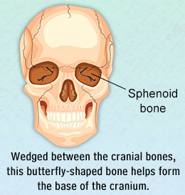 Did You Know?The sphenoid bone articulates with all the cranial bones, which is why it is referred to as the 'keystone of the cranial floor'.Out of the 206 bones found in the human body, 22 bones are found in the skull. Out of these 22 bones, 8 are the bones of the cranium, and the rest are facial bones. The bones of the cranium include the frontal bone, 2 parietal bones, occipital bone, sphenoid bone, 2 temporal bones, and ethmoid bone. The sphenoid bone has a rather interesting shape. It is referred to as Os sphenoidale in Latin. The words 'sphen' and 'eidos' mean wedge and form respectively.Wedged in the center of the skull, it looks like a bat or a butterfly, with its wings extended. One of the structurally complex bones of the human body, the sphenoid comprises a median body, two greater wings, two lesser wings, and two pterygoid plates. The main function of the sphenoid bone is that it helps in the formation of the sides of the skull, the base of the neurocranium, as well as the floors. It also helps in the formation of the walls of each of the orbits, which are the two cavities that contain the eyes. This bone lies anterior to the temporal bones and forms the base of cranium, right behind the orbitals.Sphenoid Bone Location
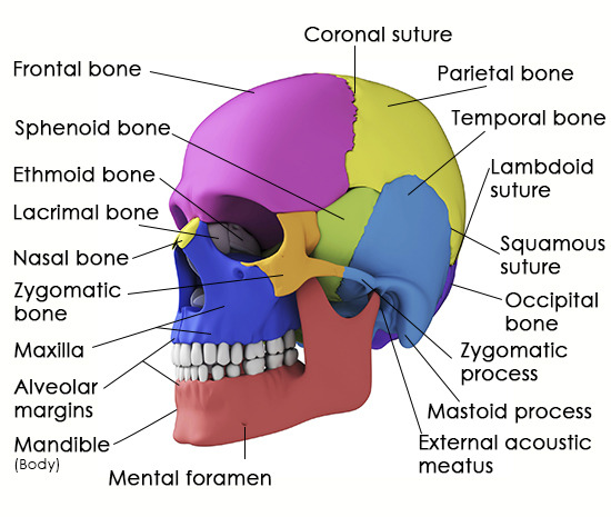 Lateral view of the skull
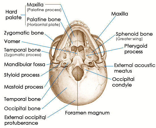 Inferior view of the skullSphenoid Bone AnatomyBesides being instrumental in the formation of the integral anatomical structures of the skull, this bone is also important as:
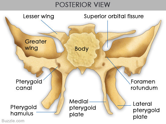 View from the back of the skullMedian BodyThe body, which is also called Corpus ossis sphenoidalis, is a cuboid-shape section of the sphenoid bone that is located at the center. In all, there are six surfaces, which include, the upper, lower, and posterior surfaces on both sides. The body contains the sphenoid sinuses, one of the four air-filled cavities located in the skull that's connected with the nasal cavity. Located on each side of the body is a carotid sulcus (canal-like passageway) for the internal carotid artery. On the upper surface of the body lies sella turcica, which is a large cavity for the pituitary gland. The sella turcica comprises the square-shaped dorsum sellae (at the back), the tuberculum sellae (at the front), posterior clinoid process, and hypophysial fossa (within sella turcica). The posterior clinoid process extends on the left and right side of the dorsum sellae. The posterior and anterior clinoid processes enclose the posterior and anterior wall of sella turcica around the pituitary gland respectively. Sphenoidal crest (narrow ridge of the bone) lies towards the front of the sphenoid bone, and sphenoidal conchae that lie on either side of the crest confine the orifice of the sphenoid sinus.
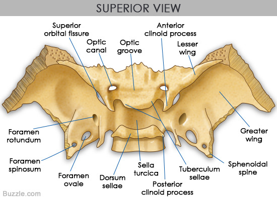 View from the top of the skullLesser WingsLesser wings, also called Ala minor, are actually the smaller of two flattened, triangular-shaped, wing-like bone plates that extend on the lateral surface on both sides of the body of the sphenoid bone. Below them lie the paired greater wings. Optic canals that lead into the orbits of the eyes are located at the base of the lesser wings. The lesser wings are a tiny section of the medial posterior wall of the orbit, and their free edges act as a border between the anterior and middle cranial fossae. The edges at the front of the lesser wings are connected with the orbital section of the frontal bone, as well as the cribriform plate of the ethmoid bone. The superior orbital fissure, which is a narrow opening located between the greater and lesser wings runs diagonally along the back of orbit. Oculomotor, trochlear, trigeminal, and abducens nerves pass through the fissure. The optic nerve and the ophthalmic artery pass through the optical canal that is located along the wings.Greater WingsGreater wing, also referred to as Ala major are larger than the lesser wings. These bone plates curve upwards, to the side and back. These assist in the formation of the floor, as well as the lateral wall of the middle cranium. They have four surfaces. While the greater wings begin with a broad base at the lateral surface of the sphenoid bone body. These wings each have four surfaces (cerebral, orbital, temporal, and maxillary). On the cerebral surface that faces the cranial cavity, lies a round opening called foramen rotundum, through which the maxillary nerve branch of the trigeminal nerve passes. It is anterior and medial to the foramen ovale, which is an oval opening that acts as a passageway for the mandibular nerve, accessory meningeal artery, lesser petrosal nerve, and emissary veins. Posterior to the foramen ovale lies foramen spinosum. The middle meningeal artery and the meningeal branch of the mandibular nerve pass through the foramen spinosum. The orbital surface forms the lateral wall of the corresponding orbit, whereas the infratemporal crest lies on the temporal surface.Pterygoid ProcessesPterygoid processes, also called Processus pterygoideus, are two bony processes that descend downwards from the junction of the greater wings and the body of the sphenoid bone. A pterygoid canal passes from back to front at the base of each pterygoid process. Each of these processes comprise a lateral and medial plate. Pterygoid fossa is a concavity or depression that lies between the lateral and medial plates. The lateral pterygoid muscle that facilitates the movement of the mandible while chewing, attaches to the lateral plate. The muscles involved in swallowing (superior pharyngeal constrictor muscle and the tensor veli palatini muscle) attach to the medial plate. A hook-shaped extension of the medial plate called pterygoid hamulus also helps in the process of swallowing.On a concluding note, the complex sphenoid bone structure is attributed to the fact that as it articulates with several cranial bones. It assists in the formation of the orbits, and also serves as an attachment of important muscles that facilitate chewing and swallowing. To add to that, it also acts as a passageway for important nerves and blood vessels.
Copyright © www.orthopaedics.win Bone Health All Rights Reserved