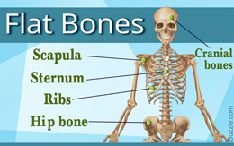
Besides some of the facial bones and cranial bones, bones such as the sternum, ribs, scapula, and the hip bone are classified as flat bones. Buzzle provides a list of all the flat bones in the human body.
Did You Know?Highest number of red blood cells in adults are found in the flat bones. These bones have a marrow, but not a bone marrow cavity.The human skeleton is a bony framework that not only gives shape to the body, but also protects the vital internal organs. It is the contraction of the skeletal muscles which are attached to the bones that facilitates movement. Moreover, the bone marrow of certain bones also produces the red blood cells and white blood cells. At birth, the human skeleton comprises about 300 bones, but the number of bones decreases to 206 in adults. The human skeleton consists of an axial skeleton and an appendicular skeleton. While the axial skeleton consists of the skull, sternum, ribs, and the vertebral column (bones that are along the imaginary longitudinal axis), the appendicular skeleton includes the bones of the arms, legs, shoulder girdle, and the pelvic girdle. The axial skeleton and the appendicular skeleton consist of 80 and 126 bones, respectively.
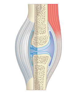
The bones of the human body are classified into long bones, short bones, sesamoid bones, flat bones, irregular bones, and wormian bones (intra-sutural). The long bones include femur, tibia, fibula, radius, ulna, and humerus. The cube-shaped short bones include the carpals, tarsals, metacarpals, metatarsals, and phalanges. Sesamoid bones are small bones that are embedded in some tendons. Patella (kneecap) is an example of a sesamoid bone. Irregular bones, as the name suggests, have an irregular shape. Hyoid bone and the vertebrae are examples of irregular bones. The flat bones are thin, as well flattened.Structure of Flat Bones in the Human BodyAs the name suggests, flat bones are strong flattened plates of bone. These are curved and have a large surface for muscle attachment. Most of them provide protection to the soft tissues or vital organs that lie under them. To understand the structure of flat bones, one needs to understand the difference between a compact bone and a spongy bone. Basically, these two types of bone tissue differ in terms of density.
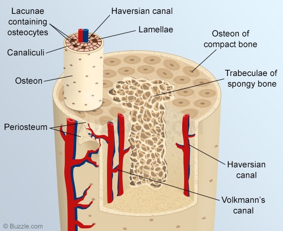
The compact bone is made up of osteons that are tightly packed. The osteons comprise an osteonic (haversian) canal, which is a central canal that contains a few blood vessels and nerve filaments that are surrounded by concentric rings of matrix called lamellae. Between these lamellae lie small chambers (lacunae) that contain osteocytes (mature bone cells) in a concentric arrangement around the osteonic canal.
On the other hand, spongy bones are less dense. They consist of trabeculae or bar-shaped bones that are arranged along the lines of stress. These provide strength to the ends of the weight-bearing bones. The spaces between them contain red bone marrow. In case of flat bones, spongy/cancellous bone lies between two layers of compact bone. The structure of these bones is such that they provide protection. In case of the skull bones, the layers of compact tissue are referred to as the tables of the skull. While the outer layer is tough and thick, the inner layer is thin, dense, and brittle. This thin layer is referred to as the vitreous table. The cancellous or spongy tissue is also called the diploë. In certain regions of the skull, the spongy tissue gets absorbed, leaving behind air-filled spaces (sinuses) between the two tables.List of Flat Bones
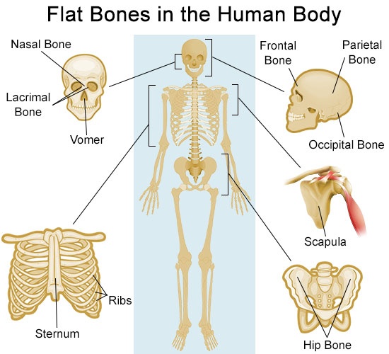
Flat bones are broad bones that provide protection or muscle attachment. These bones are expanded into broad, flat plates, as in the cranium (skull), hip bone (pelvis), sternum, rib cage, and scapula. The flat bones of the human body are as follows:
● Occipital
● Parietal
● Frontal
● Nasal
● Lacrimal
● Vomer
● Scapula
● Os coxæ (Hip bone)
● Sternum
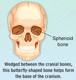
● RibsCranial and Facial BonesThe cranial bones include occipital bone, two parietal bones, frontal bone, two temporal bones, sphenoid bone, and the ethmoid bone. The top and both sides of the head are formed by the paired parietal bones. The frontal bone forms the forehead, whereas the occipital bone forms the back of the head. These are all thin, curved plates that protect the brain in the event of a traumatic injury. There are fourteen facial bones, which include the maxilla, zygoma, lacrimal, nasal, inferior nasal conchae, palatine, vomer, and mandible. Out of these, nasal bone (two oblong-shaped bones that form the bridge of the nose), lacrimal bone (smallest bone of the skull that is in the anterior part of the medial wall of the orbit), and vomer (quadrilateral-shaped bone that forms the lower and hind part of the nasal septum) come under the category of flat bones.RibsThe human rib cage comprises twelve pairs of curved flat bones called ribs, twelve thoracic vertebrae, and a T-shaped bone called sternum. The ribs are further divided into true ribs, false ribs, and floating ribs. The first seven pairs of ribs are referred to as the true ribs. The ends of these ribs are attached to the sternum through costal cartilage, which is a connective tissue. The next three pairs of ribs, that are called false ribs, only connect to the costal cartilage of the lowest pair of true ribs. The last two pairs of ribs are called floating ribs. These are only attached to the spine and are not connected to the sternum.ScapulaThe scapula, which is commonly known as the shoulder blade, is a triangular-shaped bone that forms the posterior section of the shoulder girdle. It joins the humerus (upper arm bone) with the clavicle (collarbone). It is a flat, paired bone with an extensive surface for muscular attachment. It has three angles (lateral, superior, and inferior), three borders (superior, lateral, and medial), three processes (acromion, spine, and coracoid process), and two surfaces (costal and posterior).SternumCommonly referred to as the breastbone, the sternum is a flattened T-shaped bone that is situated in the upper middle region in the anterior section of the chest. It is a part of the rib cage. It attaches to the cartilage of the true ribs (first seven pairs of the ribs) and the clavicle on either side. It is convex-shaped in the front and slightly concave at the back.Os coxæ (Hip bone)The right and left hip bones, sacrum, and the coccyx form the pelvis in the human body. The right and left hip bones meet in the front at pubis symphysis, and connect with the sacrum at the back. Each hip bone consists of 3 sections called ilium, ischium, and pubis. These three bones make up the anterolateral section of pelvic girdle. Ilium is the largest of these bones and forms the superior section of the hip bone. Ischium forms the lower section at the back, while pubis forms the lower section in the front. These bones are separate in childhood, but fuse to form the hip bone by the age of 25 years.Flat bones are important as these not only protect vital organs and tissues, but also provide a large surface area for ligament and tendon attachment. Moreover, the spongy bone tissue that is located between the layers of tough compact bone tissue also contains red bone marrow.