Anatomy of the sacrum is a bit complicated, as this bone is a fusion of five vertebrae. This article provides information related to the anatomy and details about the structure of this bone.
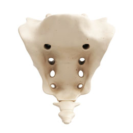
In vertebrates, the sacrum is an important bone that is present between the two hip bones. This is a large triangular-shaped bone that is present in the region of the base of the spine, forming a part of the central skeletal system. The word sacrum comes from the Latin word
sacer, meaning sacred or strong, as it was believed that this is the bone that houses the organs of procreation. It actually is a single bone, but is made by the incomplete fusion of five vertebrae, which occurs during the age of sixteen to eighteen years, and is completed by the time the person reaches twenty-five years of age.
Anatomy
The sacrum is a kyphotic bone, that is, it is concave in shape, facing outwards and forwards. There lies an intervertebral disc between the base of the last lumbar vertebra and the sacrum, which is known as the lumbosacral disc. The canal for the passage of the spinal cord extends into the sacrum. The sacral nerves exit through the bony foramina, which are present along the outer borders of the sacrum. If these nerves get pressed due to dislocation or any other injury, then it can lead to intense pain in that area, among other symptoms. The pelvic side of this bone has four separate ridges that help in demarcating the fusion of different vertebrae. Its anatomy is described below by dividing this bone into the following parts:
Sacral Ala
They are the outward and lateral winglike projections present in the superior portion of the sacrum. These have anatomical and surgical importance, as these help form the joint between the sacrum and the pelvis. When a spinal surgery is in progress, these projections are used as points of attachment for instruments, which helps to stabilize the lumbosacral joint.
Body
It is the main central portion of the sacrum that directly leads to the coccyx bone, also known as the tailbone. The central area is marked by the medial sacral crest, which slightly deviates towards the side near its end, which is known as the sacral hiatus. On the dorsal portion, there are four different converging grooves for the sacral nerves, which are laterally present to the median central crest by an inch. Behind the main body is a huge vertebral foramen, which leads straight into the sacral canal that is roofed by the fused vertebral laminae. The superior portion has two structures, known as sacral horns or superior articular processes, which are supported by short and heavy structures known as pedicles.
Sacroiliac Joint
The sacrum bone articulates with the ilium, which is the hip bone on the sides, at the point of the sacroiliac joint. The sacrum is strategically positioned between the two ilia, wherein its upper and wider part, which is known as the pars lateralis, articulates with the ilium on either side of the sacrum. The sacroiliac joints are extremely important ones, as they bear the body's weight when a person twists or turns along the sides. Their importance can be gauged by the fact that the ligaments, holding together the bones that form this joint, are among the strongest ones in the body.
The sacrum eventually leads to the coccyx, which is the terminal portion of the spine, made up of four to five fused vertebrae. It is important to know the sacrum anatomy in detail because of its significance in various surgical procedures, like correction of hip displacement and also during hip replacement procedures.