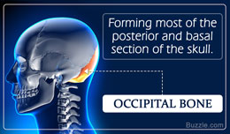
The occipital bone is a trapezoid-shaped bone that forms the posterior wall and the base of human skull. Buzzle explains the anatomy, diagram, and function of the occipital bone.
Did You Know?When we nod, we use the atlanto-occipital joint, which is the joint located between the atlas (vertebra C1) and the occipital bone. This joint helps in the flexion and extension of the neck.The term 'cranium' refers to the part of the skull that encloses the brain. Out of the 22 bones that form the human skull, 8 bones form the cranium. The cranial bones of the human body include the occipital bone, parietal bones, frontal bone, temporal bone, sphenoid bone, and the ethmoid bone. These bones perform the vital function of protecting the brain from damage in the event of trauma. There are four major sutures (fibrous joints) that join the cranial bones. These include coronal suture (suture that joins the frontal bone to the two parietal bones on each side), lambdoid suture (suture that separates the occipital bone from the two parietal bones), sagittal suture (suture that extends from the lambdoid to the coronal suture between the parietal bones), and the squamous suture (suture that forms the boundary between the parietal and temporal bone on each side).
Forming the posterior surface of the skull, the occipital bone articulates with the two parietal bones, sphenoid bone, two temporal bones, and the atlas (first cervical vertebra). The term 'occipital' is derived from the Latin word 'occiput', which in turn is derived from the prefix 'ob' (which means against) and 'caput' (which means head). The prominent occipital bone characteristics or features include the external occipital protuberance and crest, occipital condyles, and the superior and inferior nuchal lines.Occipital Bone Location and Function
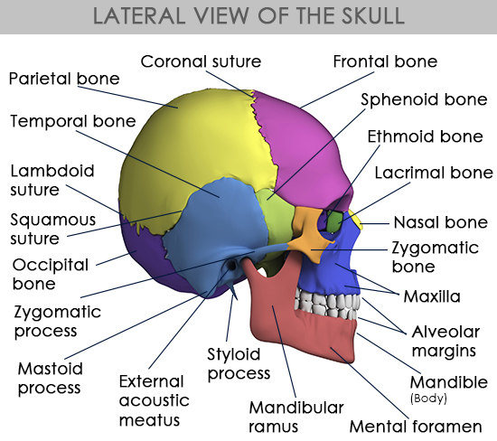
Lateral view of the skullThe occipital bone is located at the lower section of the back of the skull. It is convex-shaped externally and concave-shaped internally. It is divided into four regions. These four regions surround the foramen magnum, which is a large hole that is located at the center of the occipital bone. The four regions that are integral to the occipital bone anatomy include:
➠ Basioccipital
➠ Ex-occipitalis (two condylar sections)
➠ Squama occipitalis
At birth, the aforementioned parts develop separately, and are joined by cartilage, thereby forming a ring around the large opening called foramen magnum. It is only around the age of six years that these four parts unite to form a single bone. The squamous section, which is formed by a concave, posterosuperior plate behind the foramen magnum, makes a major part of the occipital bone. The basilar section is located in front of this foramen. The two lateral sections are located on either side of the foramen magnum.
The foramen magnum is located at the center of the occipital bone. It acts as a passageway for the spinal cord. Medulla oblongata of the brainstem enters and exits the skull through this aperture. It is here that the medulla oblongata becomes continuous with the spinal cord. Thus, it plays a vital role in the communication between the brain and the spinal cord.
Besides the medulla oblongata, meninges, subarachnoid space, the tonsils of the cerebellum, and other anatomical structures pass through the foramen. These include ligaments (apical ligament of dens, cruciform ligament of atlas, and membrana tectoria), anterior and posterior spinal arteries, vertebral artery, meningeal branches (C.N.1 to 3), sympathetic plexuses, spinal vein, spinal roots of the accessory nerve, etc.
The occipital bone provides attachment for a number of muscles of the neck, as well as the upper section of the back. These include:
➠ Longus capitis
➠ Rectus capitis anterior
➠ Rectus capitis lateralis
➠ Rectus capitis posterior major
➠ Rectus capitis posterior minor
➠ Superior oblique
➠ Semispinalis capitis
➠ Splenius capitis
➠ Occipitofrontalis
➠ Trapezius
➠ SternocleidomastoidSquama The squama, which is an expanded plate that is located above and behind the foramen magnum, is convex externally and concave internally. The major features of this region include:External SurfaceLocated on the midline of the external surface, the external occipital protuberance is a prominence that can be felt on touching. It acts as a site of attachment for the trapezius muscle. Nuchal lines are curved lines or ridges on the external surface, where muscles and ligaments of the neck and back attach to the skull. These lines form, as the stress on the bone in the areas where muscles attach to the skull causes them to develop raised bone.
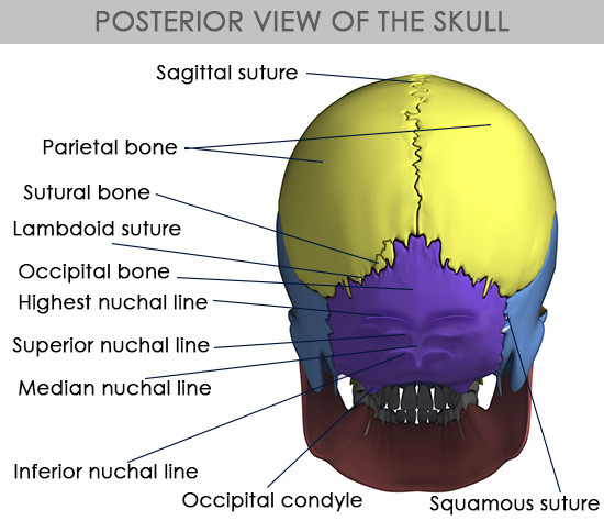
Posterior view of the skullFour nuchal lines are present on the external surface of the occipital bone. These include:
➠ The highest nuchal line, which extends on either side from the external occipital protuberance to the mastoid process of the temporal bone, is the site of attachment for galea aponeurotica (muscle that covers the upper part of the cranium).
➠ The superior nuchal line, which lies below the higher nuchal line, serves as a site of attachment for the trapezius, sternocleidomastoid, and splenius capitis muscles.
➠ The medial nuchal line, which is also called external occipital crest, begins at the external occipital protuberance and runs down to the foramen magnum. It also serves as a site of attachment for the nuchal ligament that is present at the back of the neck.
➠ Extending laterally on either side from the middle of the medial nuchal line is the inferior nuchal line that almost runs parallel to superior nuchal line. It serves as a site of attachment for the semispinalis capitis muscle.Internal SurfaceThe internal surface of the squamous part is concave in shape. The cruciate eminence divides it into four fossae (concavity in a surface). The internal occipital protuberance lies at the point of intersection of the four divisions. The upper and lower divisions are triangular and quadrilateral in shape, respectively. The upper two divisions accommodate the occipital lobes of the cerebrum, whereas the lower two divisions accommodate the cerebellar hemispheres (hemispheres of the cerebellum). At the point of intersection of the four divisions of the cruciate eminence is the internal occipital protuberance.
Grooves are present on the internal surface due to the presence of the dural venous cranial sinuses (venous channels that drain blood from the brain to the jugular foramen) such as the superior sagittal sinus, the transverse sinuses, and the sigmoid sinus. The angle of the union of the superior sagittal and transverse sinuses is called torcular herophili, and it appears as a depression on one or other side of the protuberance. Grooves for the transverse sinus run horizontally from the left and right of the internal occipital protuberance and continue into the indentations or grooves for the sigmoid sinus.
The margins of the transverse occipital sulcus serve as a site of attachment for the tentorium cerebelli (a compartment or fold created by the dura mater, which is outermost membrane that encloses the brain). The dura creates dural folds called falx cerebri and the tentorium cerebelli. Falx cerebri separates the right and left hemispheres of the brain, whereas tentorium cerebelli separates the cerebrum (the largest part of the brain in the anterior that comprises the two hemispheres) from the cerebellum (part of the brain that is situated above the medulla oblongata and beneath the cerebrum). In the upper division, to the right, is the groove that accommodates the rear part of the superior sagittal sinus. To the margins of this sulcus are attached the falx cerebri. The lower division serves as a site for attachment to the falx cerebelli. Occipital sinus lies in the attached margin of falx cerebelli. The term 'vermian fossa' refers to a small cavity or depression present in the lower part of the internal occipital crest that is occupied by part of the vermis of the cerebellum.
The occipital bone consists of the superior and inferior angles, as well as superior and inferior borders. While the superolateral borders (parietal margin) join the posterior border of the parietal bone (lambdoid suture), the inferolateral borders join with the mastoid section of the temporal bone on either side (occipitomastoid suture). At the confluence of the superolateral borders lies the superior angle. In adults, the superior angle articulates with the occipital angles of the parietal bones, whereas the inferior angle joins with the body of the sphenoid bone. The superior angle is around the lambda, located at the junction of the sagittal and lamboid sutures of the skull. On each side, where the superolateral and inferolateral borders meet, lies the lateral angle.Lateral Section/Condylar Parts
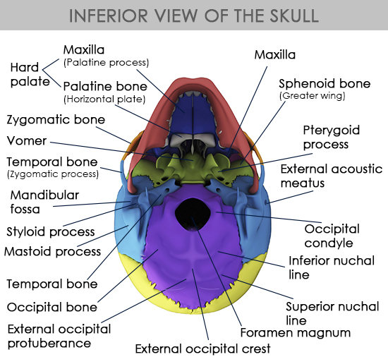
Inferior view of the skullLocated laterally to the foramen magnum on either sides, the condylar parts consist of two kidney-shaped prominences. These prominences are referred to as occipital condyles. These articulate with the atlanto-occipital joint (first cervical vertebra C1). The capsules of the atlanto-occipital articulations are attached to the margins of condyles. A bony protuberance for the attachment of the alar ligament is present on the medial side of each of these condyles. A short, bony canal called the hypoglossal canal (anterior condyloid foramen) is present at the base of each of these condyles. The hypoglossal or twelfth cerebral nerve exits through the canal, and the meningeal branch of the ascending pharyngeal artery enters through it.
Condylar fossa lies behind each condyle, and the opening for the condylar canal lies at the bottom of the fossa. The jugular notch is located on the side of the condyle. The groove for the signoid sinus lies on the cerebral surface of the bone, near the jugular notch. The emissary veins (connecting veins in the head and the scalp that drain blood from the dural sinuses to veins outside the skull) pass through these canals from the transverse sinus.
The jugular process, which is a quadrilateral-shaped plate of bone, runs sideways from the posterior half of the condyle. The rough, underside of the jugular process serves as a site of attachment for the rectus capitis lateralis muscle, as well as the lateral atlanto-occipital ligament. Also, the paramastoid process that projects from the surface might articulate with the transverse process of the atlas. A deep groove on the upper surface of the jugular process curves towards the middle and the front. It is continuous with the jugular notch. The last section of the transverse sinus is accommodated by this groove. The notch also forms the rear section of the jugular foramen.
The jugular tubercle, an oval-shaped eminence in the upper surface of the lateral section, lies above the hypoglossal canal. Glossopharyngeal, vagus, and accessory nerves pass through the oblique grooves lying in the posterior of the tubercle.Basilar PartExtending forward and upward from the foramen magnum is the basilar part. About 1 cm in front of the foramen magnum lies a pharyngeal tubercle. This tubercle serves as a site of attachment for the fibrous raphé of the pharynx. The longus capitis and rectus capitis anterior insert on either side of the middle line, whereas the anterior atlantooccipital membrane is attached right in front of the foramen magnum.
A shallow indentation or groove that extends upward and forward from the foramen magnum lies on the upper surface of the basilar part. Besides providing support to the medulla oblongata, it serves as a site of attachment to the membrana tectoria, near the margin of the foramen magnum. Small grooves for the inferior petrosal sinus are present on the lateral margins of this surface. In children, the basilar part connects to the body of the sphenoid bone by a cartilaginous plate. It is only after the age of 25 years, that this plate ossifies, and the occipital and sphenoid bone become continuous. On a concluding note, the occipital bone plays a vital role in the human body, as it not only protects the brain and supports the back of the head, but also facilitates the communication between the cranial cavity and the vertebral column through the foramen magnum that is located at its center.