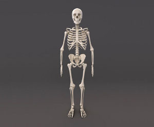
The bones provide a structural framework and protection to the soft organs. They also provide for the attachment of muscles, and help us move around.

The number of bones in the human body at birth is 300. However, as a child grows, some of the bones fuse together. The result is that there are 206 bones in the body of an adult human being. This difference in the number of bones helps forensic anthropologists in determining the age of an individual through the skeletal remains, mainly the skull. The various bones form the skeletal system, and the main function of the skeletal system is to provide a framework for the human body, and protect the delicate organs.List of Bones in the Human Body
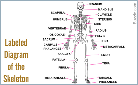
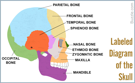
The human skull or cranium is made of 8 bones in all. It houses the brain, and forms a hard, protective covering around this master organ.Bones at a GlanceFrontal (1)
Parietal (2)
Temporal (2)
Occipital (1)
Sphenoid (1)
Ethmoid (1)
Total number of bones=8Frontal BoneThis bone forms the forehead, the roof of the orbital cavity (eye socket), and the root of the nose. A newborn has a frontal bone that consists of two parts, separated by the frontal suture. However, the parts fuse to form a single bone, by the time a child is eight years old.Parietal BonesOne bone from each side joins behind the frontal bone to form the sides and the roof of the cranium. There are 2 parietal bones, and each bone is roughly quadrilateral in shape.Temporal BonesThere are 2 temporal bones in all, one on each side below the parietal bones. The temporal bones are located lateral to the temporal lobes of the brain, and each bone consists of five parts.Occipital BoneThis is a single bone that is present at the back and lower part of the cranium, just behind the parietal and temporal bones. It has an oval aperture, known as the foramen magnum, through which the spinal cord enters the skull. Vertebral arteries, spinal nerves and ligaments that join the skull to the vertebrae, pass through this aperture.Sphenoid BoneThis is a single bone that is situated at the base of the skull in front of the temporal bones and basilar part of the occipital bone. It's shape resembles that of a butterfly, and it is one of the seven bones that form the orbital cavity or eye socket.Ethmoid BoneThis is a light and spongy bone that is situated in the anterior part of the base of the cranium. It lies between the two orbits, at the roof of the nasal cavity, separating the brain from the nasal cavity. It is one of the seven bones that form the orbital cavity, and consists of three parts.Facial Bones at a GlanceMandible (1)
Maxilla (2)
Palatine bone (2)
Zygomatic bone (2)
Nasal bone (2)
Lacrimal bone (2)
Inferior nasal conchae (2)
Vomer (1)
Total number of bones=14MandibleThis is the lower jawbone, and is known as the inferior maxillary bone. It is U-shaped and is the largest and strongest bone of the face. The mandible of a newborn consists of two halves that fuse at the mental symphysis during the first year. Each half of the mandible has a horizontal body and a vertical ramus at the posterior end of the body. The part of the mandible that bears teeth, is known as the alveolar process.MaxillaThe maxilla, or the upper jawbone, holds the teeth of the upper jaw, and forms the walls of the orbital cavity. It also contributes to the roof of the oral cavity, and the lateral walls and floor of the nasal cavity. The mandible is actually two bones that are fused along the palatal fissure. Failure of fusion of the two bones before birth, can lead to a congenital deformities, such as cleft palate(palatoschisis) and cleft lip (cheiloschisis).Palatine BoneThe palatine is an L-shaped bone that is situated between the maxilla and the pterygoid process of the sphenoid. It is located behind the nasal cavity and hence, contributes to its floor and the lateral walls. Besides the nasal cavity, it also contributes to the roof of the mouth as well as to the floor of the orbit.Zygomatic BoneThe zygomatic bone is also known as the cheekbone or malar bone. There are 2 such bones, one on each side of the face, forming the prominence of the cheek. It is one of the many bones that form the walls of the orbital cavity.Nasal BoneThe nasal bones are two in number, and together form the bridge of the nose. These are 2 oblong bones, and their size varies in different individuals.Lacrimal BoneThe lacrimal bone is the smallest bone of the face, and there are 2 such bones, each one forming a part of the median wall of the orbital cavity. Each lacrimal bone articulates with the frontal bone, the ethmoid bone, the maxilla, and the inferior nasal concha.Inferior Nasal ConchaeThese are paired bones of the face that arise from the maxillary bone and continue horizontally along the lateral wall of the nasal cavity. Located above these bones, are the middle nasal concha and the superior nasal concha, which arise from the cranial region.VomerIt is a thin, flat single bone that lies along the midsagittal line. It articulates with 6 bones: the ethmoid, the sphenoid, the two palatine bones, and the two maxillary bones.
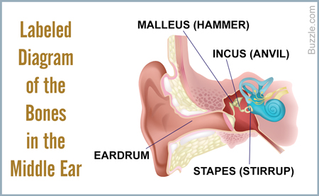
The middle ear is the region between the eardrum and the cochlea. In all mammals, including humans, the middle ear has three bones that are known as the auditory ossicles. These are very small bones, and their function is to transmit sound vibrations from the eardrum to the inner ear.Bones at a GlanceMalleus (2)
Incus (2)
Stapes (2)
Total number of bones=6Malleus (hammer)The malleus is a small hammer-shaped bone that is attached to the eardrum at one end, and to the incus at the other. Its function is to transmit sound vibrations from the eardrum to the incus. The malleus is only found in mammals.Incus (anvil)The incus is an anvil-shaped bone that connects the malleus to the stapes. Its function is to transmit sound vibrations from the malleus to the stapes.Stapes (stirrup)The stapes is a stirrup-shaped small bone that is attached to the incus at one end, and to the fenestra ovalis or the "oval window", on the other. The oval window is nothing but an opening to the inner ear, and it is covered with a membrane. The function of the stapes is to transmit sound vibrations from the incus to the labyrinth of the inner ear. It is the smallest bone in the human body.Bones of the Shoulder GirdleThe shoulder is made of two bones that together allow the attachment of the arm to the body. The bones of the shoulder are as given below. Bones at a GlanceScapula (2)
Clavicle (2)
Total number of bones=4ScapulaThe scapula is a flat, triangular bone that forms the posterior part of the shoulder girdle. It connects the humerus (upper arm) with the clavicle. It is commonly referred to as the shoulder blade. There are two such bones, one on each side of the shoulder.ClavicleIt is commonly called the collarbone, and is a pair of small long bones that join the scapula to the sternum. Put in simple words, it is the bone that attaches the arm to the body. An interesting fact about the clavicle is that it is the only long bone that lies horizontally.Bones of the ThoraxThe thorax is the part of the body between the neck and the abdomen. It is the portion below the neck that encloses the heart and the lungs. Here's a list of the bones in the thorax.Bones at a Glance Scapula (2)
Clavicle (2)
Total number of bones=4SternumThis is a long T-shaped bone. It lies in the central portion of the rib cage, and is attached to the ribs via cartilage. Together with the ribs, it forms the anterior part of the rib cage. It consists of three parts, and the topmost part is the manubrium, to which the clavicle is attached. This is followed by the body (to which the ribs are attached), and the xyphoid process.RibsThere are 12 pairs of ribs in all. The first seven pairs are directly attached to the sternum through cartilage. The next three are attached to the sternum through a common cartilaginous extension. The last two pairs are known as the floating ribs because although they start from the thoracic vertebrae, they do not attach to the sternum.
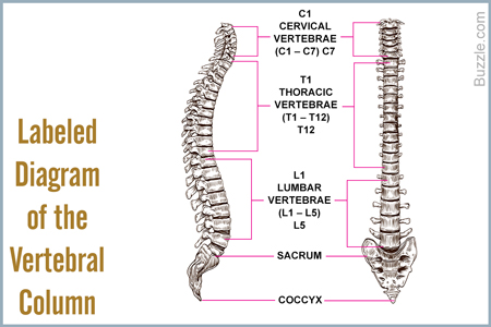
The vertebral column, or the spinal column, is made up of 24 small bones, each of which is known as a vertebra. In addition to the vertebrae, there are two other bones that form a part of the vertebral column: the sacrum and the coccyx. The vertebrae give the vertebral column its flexibility due to which we can bend forwards and sideways. The various vertebrae are grouped as given below.Bones at a GlanceCervical vertebrae (7)
Thoracic vertebrae (12)
Lumbar vertebrae (5)
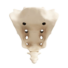
Sacrum (1)
Coccyx (1)
Total number of bones=26Cervical VertebraeThe cervical vertebrae are the first 7 vertebrae that are also the smallest of the true vertebrae. They are different from those in the thoracic and lumbar regions for the fact that they have a hole or foramen in each transverse process for the vertebral artery to pass through. The skull is supported by the first cervical vertebra, which is known as the atlas. The second cervical vertebra, known as the axis, forms the pivot on which the atlas turns. The cervical vertebrae form the neck. The cervical vertebrae are designated as C1 to C7, as shown in the diagram.Thoracic VertebraeThe thoracic vertebrae form the middle region of the vertebral column, and are located between the cervical and the lumbar vertebrae. There are 12 thoracic vertebrae, which are larger than the cervical vertebrae but smaller than those in the lumbar region. The distinct features of these vertebrae are the presence of facets for the attachment of ribs. Each thoracic vertebra has facets on the side of the bodies, except the last two. The thoracic vertebrae are designated as T1 to T12, from top to bottom.Lumbar VertebraeThe lumbar vertebrae consists of 5 vertebrae, located in the region between the ribs and the pelvic girdle. They lack the foramen on transverse processes that characterize the cervical vertebrae, and also the facets on the body that are the distinct features of the thoracic vertebrae. The lumbar vertebrae are designated as L1 to L5, from top to bottom.SacrumFive sacral vertebrae fuse to form a triangular bone called the sacrum in adults. The sacrum fits between the two hip bones and joins the spine and the pelvis together. The two lateral projections of the sacrum articulate with the ilium.CoccyxThe coccyx is referred to as the tailbone, and consists of 4 bones that fuse together as one grows up. Coccyx can variably consist of 5 or 3 bones as well. It is attached to the base of the sacrum by a fibrocartilaginous joint. The coccyx is a remnant of a vestigial tail in all tailless primates.
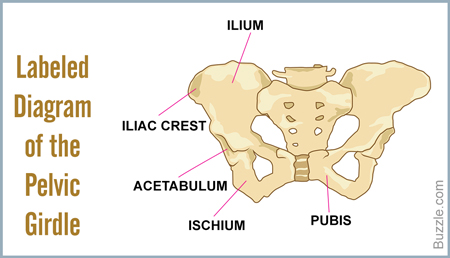
Bones at a GlanceHip bone (2)
Total number of bones=2This is an irregularly shaped bone that is constricted in the middle and flared at both ends. There are two hip bones that join together to form the pelvic girdle or pelvis. Each hip bone has three parts - the ilium, the ischium and the pubis.The ilium is the flared, fan-shaped superior portion of the hip bone. The ischium is the lowest portion of the hip bone that curves forward and meets the pubis to form the obturator foramen.
The bones of the hands can be divided into those that make up the upper arm, the lower arm, the wrist, the palm and the fingers. Bones at a GlanceHumerus (2)
Radius (2)
Ulna (2)
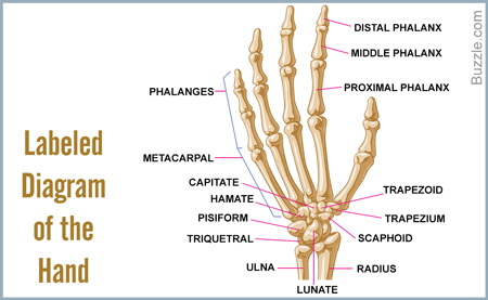
Carpals (16)
Metacarpals (10)
Phalanges (28)
Total number of bones=60HumerusThis is a single long bone of the upper arm. It runs from the shoulder to the elbow. The humerus connects the scapula to the bones of the forearm.RadiusRadius is one of the long bones of the forearm that lies on the lateral side of the ulna (the other bone of the forearm). Prismatic in shape, it starts from the lateral side of the elbow and continues to the thumb side of the wrist.UlnaIt is a long bone that runs parallel to the radius, along the forearm. It has a prismatic shape, and lies on the side of the little finger.Carpal BonesThese are the bones of the wrist. There are 16 carpal bones in all, 8 on each wrist.
Scaphoid bone (2)
Lunate bone (2)
Triquetral bone (2)
Pisiform bone (2)
Trapezium (2)
Trapezoid bone (2)
Capitate bone (2)
Hamate bone (2) Metacarpal BonesThese are the bones of the palm, and there are 5 metacarpal bones in every palm, one corresponding to each digit.Phalangeshese are the bones of the fingers. There are 5 proximal phalanges in each hand, and they are located in front of the metacarpals. They are 4 intermediate phalanges in front of the proximal phalanges, one on each finger, except the thumb. The last phalanges are located at the tip of each finger, and are known as the distal phalanges. They are 5 in number.
The bones of the legs are those that make up the thigh, the lower half of the legs, and the feet.Bones at a GlanceFemur (2)
Tibia (2)
Fibula (2)
Patella (2)
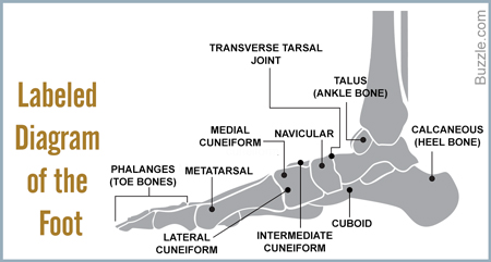
Tarsals (14)
Metatarsals (10)
Phalanges (28)
Total number of bones=60FemurThis is the longest bone in the human body, and is also known as the thigh bone. The head of the femur forms articulates with the acetabulum of the pelvic bone, at the hip joint. Its other end articulates with the patella and the tibia at the knee joint. In four-legged vertebrates, the femur is found only in the hind legs!TibiaThe tibia is the second longest bone in the human body. Along with the fibula, it forms the lower part of the leg below the knee. It articulates with the femur (thigh bone) at its superior end, and with the talus (ankle bone) at its inferior end. Laterally it articulates with the fibula. The tibia is considered by many to be the strongest bone of the body. It is commonly known as the shin bone.FibulaThe fibula is a long but thin bone which, along with the tibia, forms the lower part of the human leg. It is attached to the tibia at both the ends. Its upper end articulates with the tibia at the back of its head, whereas while attaching to the tibia with its lower end, it angles slightly forward. The fibula is also known as the calf bone.PatellaThe patella is a triangular bone that forms a protective cap over the knee joint. Also known as the kneecap, it articulates with the femur (thigh bone). It is the largest sesamoid bone in the human body.Tarsal BonesThe tarsal bones are the bones of the ankle, and there are 14 tarsal bones, 7 on each foot. They are as under.
Calcaneus (2)
Talus (2)
Navicular bone (2)
Medial cuneiform bone (2)
Intermediate cuneiform bone (2)
Lateral cuneiform bone (2)
Cuboid bone (2)Metatarsal BonesThere are 5 metatarsal bones in each foot, one corresponding to each digit. These lie between the tarsal bones and the phalanges. These may be considered to be equivalent to the metacarpal bones of the hands.PhalangesThese are bones of the toes of the feet. There are 5 proximal phalanges in each foot (as shown in the diagram above). There are 4 intermediate phalanges, one on each finger, except the big toe. The phalanges on the tips of the toes are known as the distal phalanges. They are 5 in number.In addition to the bones mentioned above, there is another bone. It is the hyoid bone which is located in the throat. It is a small bone that is attached to the muscles of the mouth, tongue, larynx, pharynx, and the epiglottis. This brings the total count of bones to 206!