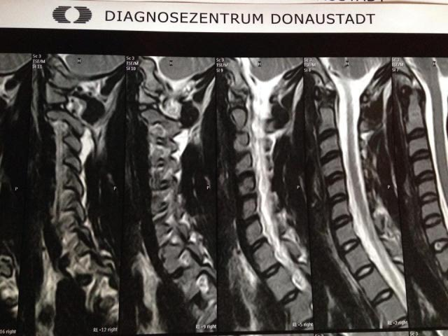
Questionhi my mum has been in and out of hospitals for the past two weeks with chronic back pain. she has been unable to sleep, sit or even use the toilet. We finally were able to get her in for an mri and the results have just come back but we are unclear about what this means for her.
would you be able to please explain what these results mean, what comes next for her and tell us possible treatments especially if she will need surgery.
Any suggestions about pain management are also appreciated.
results:
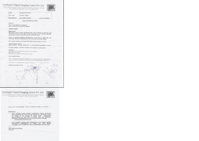
clinical history: long history of severe low back pain, prolapsed l3/4, l4/5 discs.
findings: the intervertebral discs from t11-l3 were intact and demonstrated normal morphology and signal intensity. there is no loss of disc height, desiccation, annulus tear or disc protrusion. the spinal canal and foramina are widely capacious at these levels and there is no cord compression or nerve root impingement.
l3/4: there is moderate desiccation and mild displaced narrowing. there is prominent posterior midline and left paracentral disc protrusion with a large inferiorly extruded disc fragment. this measures 8.5mm AP x 10 mm transverse x 13mm superior/inferior diameter. the disc fragment has migrated inferiorly into the left lateral recess medial to the left pedicle of l4. there is marked compression of the proximal left l4 nerve root. there is no lateral disc protrusion or impingement upon the l3 nerve roots within the foramina.
l4/5: there is mild desiccation and minimal loss of disc height. there is mild posterior midline disc annulus bulging with an associated annulus tear. the disc bulge causes mild indentation upon the anterior aspect of the dural sac and the left subarticular recess but there is no associated nerve root compression.
comments:
1. at l3/4 there is prominent left paracentral disc protrusion with a large inferior extruded disc fragment to the level of the left lateral recess causing marked compression of the proximal left l4 nerve root
2. at l4/5 there is mild degenerative with a mild degree of posterior midline disc bulging. there is no associated nerve root compression.
thank you for your time.
AnswerThis report describes degenerative disc disease. First, the disc begins to dry out which is called desiccation. Second, as it gets drier and drier it loses height which narrows the channels or holes from which exit the spinal nerve roots. This narrowing is called spinal stenosis. Third, the resultant stress on the joint causes calcium to build up. This build up has a variety of names dependent upon where it occurs and the extent of damage but in the end it is known as arthritis. This process is counter-intuitive for you would expect that wear and tear on a spinal joint would result in a wearing away of tissue, but in fact bone behaves more like skin. Under stress skin builds up into what is known as a callous, bone builds up into what are known as calcium deposits or sclerosis which gradually become bone spurs or osteophytes, in short arthritis. As the disc gets drier and drier the outside walls (annular fibers) get weaker and weaker and eventually bulge or protrude. This protrusion if large enough or in a vulnerable place will compress or pinch the spinal cord or spinal nerve root. A picture is worth a thousand words so if you'll click on
YouTube you'll see a video which will make this much clearer.
The worrisome part of this MRI is the " large inferiorly extruded disc fragment". This means that it appears as though a piece of the protruded disc (the red bubble seen in the video above) has actually broken off. Like a ball bearing loose in a transmission it will sometimes grind the gears and sometimes not depending upon its precise position. Sometimes smaller fragments can be re-absorbed by the body which treats it as a foreign body, but this fragment seems to be fairly large. With what little information I have this would appear to be a surgical case.
AUTHOR BIOGRAPHY
Dr. Michael L. Hall, D.C. practices at Triangle Disc Care in Raleigh, North Carolina specializing in Spinal Decompression for the treatment of acute and chronic neck pain and back pain due to herniated, degenerated discs. This is a conservative procedure for patients suffering with bulging or herniated discs, degenerative disc disease, posterior facet syndrome, sciatica, failed back surgery syndrome, and non-specified mechanical low back or neck pain.
For more information call 919-571-2515, click on
www.triangledisc.com or email
[email protected] . Type "Free eBook - 101 Things I Need to Know about my Bad Back" into the subject line.