The stress fracture, first described by Breithaupt in 1855,[1] is a common overuse injury seen in athletes and military recruits.[2, 3] The injury is usually seen in the lower extremities, but it has also been reported in the upper extremities and the ribs. The most common locations for stress fractures include the tibia, metatarsals, fibula, and navicular bones; less common locations include the femur, pelvis, and sacrum.
A stress fracture is caused by repetitive and submaximal loading of the bone, which eventually becomes fatigued and leads to a true fracture. The typical presentation is a complaint of increasing pain in the lower extremity during exercise or activity. The patient's history usually reveals a recent increase in either training volume or intensity.
The treatment of most stress fractures is relatively straightforward and includes decreased activity and immobilization; however, patients with some stress fractures, such as displaced femoral neck stress fractures and fifth metatarsal base stress fractures, are more likely to have complications such as nonunion.[4, 5] These complications should be monitored closely because surgical intervention may be necessary.
NextStress fractures result from recurrent and repetitive loading of bone. The stress fracture differs from other types of fractures in that in most cases, no acute traumatic event precedes the symptoms.
Normal bone remodeling occurs secondary to increased compressive or tensile loads or increased load frequency. In the normal physiologic response, minor microdamage of the bone occurs. This is repaired through remodeling. Stress fractures develop when extensive microdamage occurs before the bone can be adequately remodeled.[6, 7]
Often, the patient has a history of an increase or change in the character of activity or athletic workouts. Bones may be more prone to stress fractures if the bone is weakened, as in individuals with osteoporosis or those in whom weightbearing activities are increased.
The three factors that can predispose an individual to the development of stress fractures are as follows[8] :
The applied load on the bone may be increased by decreasing the surface area that the weight is distributed across or by increasing the total weight applied to the bone. High-impact activities, such as jumping or performing plyometrics, running on a new surface, or practicing incorrect biomechanical movements or techniques, may increase the risk of stress fractures.
Although stress fractures result from repeated loading, the exact contribution of training factors (eg, volume, intensity, and surface) has not been clearly established.[9] From what we do know, menstrual disturbances, caloric restriction, decreased bone density, muscle weakness, and leg-length differences are risk factors for stress fractures.[10, 11]
Myburgh reported that stress fractures were more common in athletes who had decreased bone density, lower dietary calcium intake, current menstrual irregularity, and less oral contraceptive use, when the athletes were matched for similar training volume and intensity.[12]
Nattiv and Armsey found that genetics, female sex, white ethnicity, low body weight, lack of weightbearing exercise, intrinsic and extrinsic mechanical factors, amenorrhea, oligomenorrhea, inadequate calcium and caloric intake, and disordered eating were additional risk factors for stress fractures.[13] A decreased testosterone level in male endurance athletes has also been implicated as a risk factor for stress fractures.[14, 15, 16, 17]
Schnackenburg et al did a matched control study on 19 female athletes with tibial stress fractures and found that stress fracture patients had lower tibial cross-sectional areas, lower trabecular bone mineral densities, and less cortical area, as well as decreased knee extension strength. They suggested that impaired bone quality of the posterior cortex and decreased muscle strength were associated with stress fractures in female athletes.[18]
Giladi identified two anatomic risk factors in military recruits. Recruits with stress fractures had significantly narrower tibiae and increased external rotation of the hip. These two variables were independent and cumulative, and when both risk factors were present, the stress-fracture morbidity was 45%.[19]
Particular locations of stress fractures are commonly associated with particular activities (see Table 1 below).
Table 1. Epidemiologic Features of Stress Fractures Based on Location and Activity (Open Table in a new window)
Location of Fracture Activity Involved Metatarsals, general Football, basketball, gymnastics, ballet, military training[20] Metatarsal, base of the second Ballet Metatarsal, fifth Tennis,[21, 22] ballet Sesamoids of the foot Running, ballet, basketball, skating Navicular Basketball, football, running Talus Pole vaulting Calcaneus Military drills, running, aerobics Fibula Running, aerobics, ballet, race-walking Tibia Running sports, dancing, ballet Patella Running, hurdling Femoral neck Distance running, military training[23] Pubic rami Military drills, distance running Pars articularis Gymnastics, ballet, cricket, volleyball, diving, football Chest, ribs Swimming,[24] golf,[25] rowing[26] Sternum Wrestling[27] Ulna Racquet sports, volleyball Olecranon Baseball, throwing sportsStudies of US military recruits revealed a higher percentage of stress fractures in female recruits than in male recruits.[2, 28, 29, 30] Bennell et al also found a 45% incidence of stress fractures in competitive female runners.[6] The women most at risk for stress fractures were those who restricted their food intake and those who had dysmenorrhea.
The triad of disordered eating, amenorrhea, and osteoporosis may be extremely prevalent in female distance runners and ballet dancers, as well as in other female athletes who believe that a low body weight or body-fat percentage provides a competitive advantage.[31, 32]
The amenorrheic female athlete may experience a prolonged state of estrogen deficiency similar to that of a postmenopausal woman. The lower estrogen levels are associated with decreased bone density; even if normal menses returns, this bone loss may be irreversible in high school – and college-aged female athletes. Early identification of female athletes who are likely to develop the female athlete triad is important for the prevention of stress fractures and for maintaining overall future bone health.[33]
In a study of military recruits, Markey found no difference in the incidence of stress fractures between recruits of various racial backgrounds.[34]
With stress fractures, the typical complaint is that of an insidious onset of pain with activity or a complaint of pain in the affected extremity with repeated loading. Usually, the patient has no recent history of trauma to the affected area.
The pain subsides at rest, but symptoms return when the patient resumes the original activity. Local tenderness and swelling are often found at the fracture site. Early diagnosis is usually based on clinical findings, and several weeks may be required before the fracture site or new bone formation is visible on radiography.
The pain may occur only with weightbearing, and it may be reproducible with continued or prolonged activity. Some patients may associate a change in training equipment or training methods with the onset of symptoms.
The common findings on physical examination may include tenderness or pain on palpation or percussion of the bone. Erythema or edema may be present at the site of the stress fracture. Loading or stress of the affected bone may also produce symptoms.
Differential diagnoses for stress fractures are varied and depend on location, symptoms, history, and physical examination.
Shin splints (medial tibial stress syndrome) are a common complaint of midtibial pain, especially in runners. While the complaints of pain are similar, stress fractures of the tibia may be differentiated from shin splints based on the history and physical examination findings.
Most athletes report a crescendo-type pain with stress fractures, as the pain increases through individual workouts and from one workout to the next. Shin splint pain tends to be present at the start of activity in those athletes who are symptomatic. Physical examination of an athlete with shin splints should reveal tenderness to palpation over a wide region of the tibia and the tibialis muscle, whereas the pain from stress fractures tends to be localized to a specific area on the tibia.
Contusions typically present with ecchymosis and swelling, as well as a history of a traumatic injury.
True fractures also tend to have an obvious history, with a traumatic event being recalled by the patient with acute onset of pain.
Muscle strains may be acute or chronic. Chronic muscle strains can be differentiated from stress fractures by the location and by factors that exacerbate or worsen the injury.
Costochondritis or rib pain may mimic the pain seen in stress fractures of the ribs. A high index of suspicion should be maintained for rib stress fractures in athletes who participate in rowing sports, such as crew rowing. The pain of costochondritis may be more diffuse or widespread than the pain from stress fractures of the ribs; however, multiple stress fractures of the ribs can occur.
Exertional compartment syndrome is most commonly seen in the lower extremities. A history of swelling in the legs with athletic activity may indicate the presence of a compartment syndrome. Increased intracompartmental pressures measured with a catheter transducer would suggest compartment syndrome.
Nerve entrapment syndromes can also mimic stress fractures, but with entrapment syndromes, numbness is also often a complaint.
Superficial peroneal nerve injuries, such as the following, should be considered:
Popliteal artery entrapment syndrome is another cause of lower extremity pain. The typical history is one of increased pain or swelling with exercise. The pain tends to be more diffuse than the pain associated with stress fractures. Measurement of ankle blood pressures before and during exercise or an angiogram may help with the diagnosis.
Morton neuroma is an irritation of one of the interdigital nerves of the foot that is often seen in long-distance runners. The pain is usually localized just proximal to the second, third, or fourth toes, and it is worse when the forefoot is squeezed from the lateral and medial sides.
Metatarsalgia presents as foot pain and may be mistaken for a stress fracture of the metatarsals. The pain of metatarsalgia usually resolves quicker than the pain from a stress fracture once treatment is initiated. Bone scans may show diffuse uptake, but they do not usually show the significant uptake seen with stress fractures. Freiberg infarction (necrosis of the second metatarsal epiphysis in female adolescents) should be considered.
Stress fractures may not show up on radiographs for the first 2-4 weeks after injury. (See the image below.) The first radiographic finding may be a localized periosteal reaction or an endosteal cortical thickening. The low sensitivity of radiography for stress fractures makes bone scanning, magnetic resonance imaging (MRI), and computed tomography (CT) the preferred tests for diagnosis.
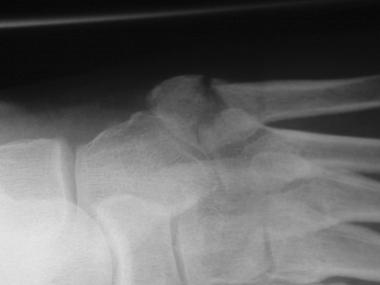 Fifth metatarsal stress fracture.
Technetium-99m bone scan findings may be positive in the case of a stress fracture after 72 hours; however, a positive bone scan finding is nonspecific, and it may be indicative of another diagnosis, such as an infection or a neoplastic process. Conventional radiography and bone scanning have been compared for the initial detection of stress fractures; positive findings were reported in 96% of bone scans, whereas only 42% positive findings were reported on radiographs.
MRI is also useful in the diagnosis of stress fractures. It provides information about bone integrity and fracture orientation, and it can demonstrate focal tissue damage and edema.
The MRI findings of stress fractures typically follow one of two patterns. In the first pattern, a hypointense, bandlike fracture line is visible with surrounding bone or tissue edema. The second MRI pattern represents an amorphous stress fracture or response pattern. In this pattern, there is no obvious fracture line or band; instead, the fracture may have diffuse or scattered areas of hypointensity on T1-weighted images, with increased signal intensity on T2-weighted images.[35]
Short-tau inversion recovery (STIR) imaging sequences suppress the normal fat signal intensity in bone marrow, allowing better visualization of intramedullary bone.[36, 37, 38]
Wright et al carried out a systematic review of published studies with the aims of assessing the diagnostic accuracy of imaging modalities used to diagnose lower-extremity stress fractures and formulating evidence-based recommendations for clinical practice.[39] Reported sensitivities and specificities (with 95% confidence interval) for these modalities were as follows:
The investigators found MRI to be the most sensitive and specific imaging test for diagnosing stress fractures of the lower extremity.[39] They concluded that when MRI is available, nuclear scintigraphy is not recommended, because of its low specificity, high dosage of ionizing radiation, and other limitations. They also noted that radiography is likely to yield false- negative results at initial presentation, particularly in the early stages of fracture, and sometimes fails to identify an existing stress fracture at any time.
Several grading systems for stress fractures based on MRI or scintigraphic findings have been proposed for correlating imaging findings with clinical findings and providing treatment guidelines (see Table 2 below).[36]
Table 2. Grading of Stress Fractures on Basis of Radiologic Findings[36] (Open Table in a new window)
Grade Radiographic Finding Bone Scan Finding MRI Finding 1 Normal Poorly defined area Increased activity on STIR image 2 Normal More intense Poor definition on STIR and T2-weighted images 3 Discrete line Sharp area of uptake No focal or fusiform cortical break on T1- and T2-weighted images 4 Fracture or periosteal reaction More intense localized transcortical uptake Fracture line on T1- and T2-weighted images
Most stress fractures can be treated conservatively by having patients stop or significantly decrease their activity for approximately 4-6 weeks, then gradually return to activity.[40, 41] (See Table 3 below.) Patients who experience pain with walking may be placed in a short leg cast with crutches, a walking boot, or a brace for 4-6 weeks. The use of pneumatic braces in the treatment and rehabilitation of tibial stress fractures also speeds the patient's return to training.[42]
Table 3. Healing Times for Various Stress Fractures*[40] (Open Table in a new window)
Site of Stress Fracture Percentage Healed at 2-4 wk, % Percentage Healed at 1-2 mo, % Percentage Healed at > 2 mo, % Tibia, proximal third 0 43 57 Tibia, middle third 0 48 52 Tibia, distal third 0 53 47 Fibula 7 75 18 Metatarsals 20 57 23 Sesamoids 0 0 100 Femur, shaft 7 7 86 Femur, neck 0 0 100 Pelvis 0 29 75 Olecranon 0 0 100 *Adapted from Hulkko.[41] Findings were from a case series of 368 stress fractures in athletes, in which the healing times of stress fractures in different locations were assessed.Nutritional measures
Regarding fracture risk, Schwellnus and Jordaan found no benefit with calcium supplementation (500 mg/day) beyond the usual dietary intake in male military recruits.[43]
Biomechanical measures
The use of orthotic devices and shoe inserts has been studied as a preventive measure for lower-extremity stress fractures. Finestone and Milgrom both studied the use of semirigid orthoses, soft orthoses, or both in the boots of military recruits during basic training.[44, 45]
Finestone found that the incidence of lower-extremity stress fractures was lower in the group using semirigid orthoses (15.7%) or soft biomechanical orthoses (10.7%) than in the control group (27%).[44] Additionally, the recruits better tolerated the soft biomechanical orthoses than the semirigid orthoses.
In a prospective study of stress fractures, Milgrom et al studied the hypothesis that a shock-absorbing orthotic device worn within military boots decreases the incidence of stress fractures.[45] Milgrom et al demonstrated a statistically significant decrease in the incidence of femoral stress fractures in the orthotic device group. In military recruits who did develop stress fractures, the time of onset and the location of stress fractures did not differ between the orthotic device group and the non–orthotic device group.
Gillespie and Grant reviewed the use of shock-absorbing insoles in four trials.[42] These insoles appeared to reduce the incidence of stress fractures and stress reactions of bone; however, incomplete data from one trial indicated that a reduction in the running distance and intensity may also have been a factor in preventing stress fractures.
High-risk stress fractures
Nonunion of stress fractures is uncommon, but it can occur. These stress injuries should be closely followed up for early surgical intervention. They include stress fractures of the neck of the femur, the anterior cortex of the tibia, the tarsal navicular, and the bases of the second and fifth metatarsals.[40]
Other high-risk stress fractures include stress fractures of the patella and medial malleolus.[46] Anterior-cortex stress fractures of the tibia are considered high-risk because the tensile forces across the anterior portion of the tibia can typically lead to delayed union or nonunion.
Low-risk stress fractures
Low-risk stress fractures include most upper-extremity stress fractures, with the possible exception of fractures through the physis of the humeral head (little leaguer's shoulder) and fractures through the medial epicondyle (little leaguer's elbow), which may have complications due to the involvement of the growth plate.
Other low-risk stress fractures include stress fractures of the ribs, pelvis, femoral shaft, fibula, calcaneus, and metatarsal shafts.
Copyright © www.orthopaedics.win Bone Health All Rights Reserved