QuestionI'm a 28 year old Female who was in a motor vehicle accident in 2003, I was diagnosed with a C1 Fracture and Occipital Condyle fracture L4/L5 fracture. The fractures didn't require surgery I wore a hard neck brace for about 12 weeks. Since then I've had chronic neck pain headaches and low back pain.
I had and MRI last year of my lower back which indicated the following
T-12-L1 Small posterior bulging disc
L1-2 Small posterior bulging disc
L2-3 Mild posterior bulging disc
L3-4 Mild posterior bulging disc. Early bilateral joint facet degeneration
L4-5 Mild/moderate borad-based posterior bulging disc slightly larger on the left. Ligamentum flavum hypertroyphy and early joint facet degeneration
L5-S1 Right paracentral posterior herniated disc and annular tear. Small extruded disc material is present adjacent to the posterior-superior aspect of the S1 vertebral body. Ligamentum flavum hypertrophy and mild joint facet degeneration
I also recently had xray's of my cervical spine which indicated the following.
1. Loss of normal cervical lordosis. This may be present with spasm.
2. There is old dens fracture present
3. Mild degenerative change involving the left neuroforamen at the C5-6 level.
After these xrays I had a cervical MRI done. Which came back normal. So could you please break this down for me and explain what exactly is wrong with me according to the MRI/Xrays. I am also curious how my cervical xrays could say one thing and my MRI be normal, and if I was diagnosed in 2003 with a C1 fracture but have an old dens fracture present? Isn't the dens considered the C2? Also any advice on treatment and or pain management. I thank you so much for your time regarding these issues.
AnswerAmy,
I will reply between lines. But I still suggest you get to a chiropractor who employs spinal decompression for your lower back and hopefully that same chiropractor will also employ "instrument adjusting" such as activator, pro adjuster or arthrostim adjusting systems those are three separate types of instrument adjusting. So when you seek a chiropractor they need to employ or use spinal decompression and instrument adjusting.
The amount of trauma you have endured is quite a lot and that is why I am recommending this type of care for our spine.
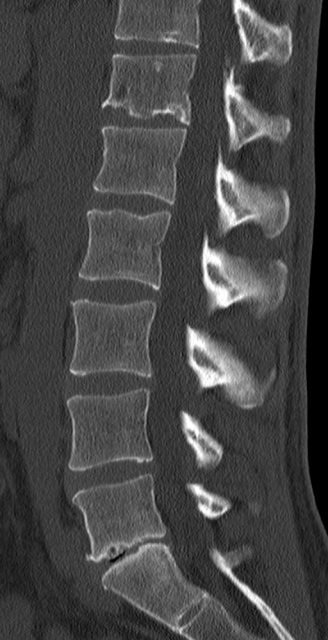
I had and MRI last year of my lower back which indicated the following
T-12-L1 Small posterior bulging disc
L1-2 Small posterior bulging disc
L2-3 Mild posterior bulging disc
THESE ARE BULGES OF THE DISC MATERIAL BETWEEN THE VERTEBRA AT THE LEVELS AS THEY ARE MENTIONED. THINK OF AN ICE CREAM SANDWICH THAT IS SLIGHTLY DEFROSTED AND YOU PUSH DOWN ON THE SANDWICH VERY LIGHTLY AND THE ICE CREAM BULGES OUTWARD. A MILD BULGE WOULD BE LESS OF AN ICE CREAM BULGE THAN MILD/MODERATE OR MODERATE BULGE.
L3-4 Mild posterior bulging disc. Early bilateral joint facet degeneration
THIS IS EXPLAINED AS ABOVE. FACET IS THE JOINT IN THE SPINE AND THEY ARE DEGENERATING MEANING THEY ARE GETTING A ROUGHENED EFFECT RATHER THAN BEING A SMOOTH SURFACE.
L4-5 Mild/moderate borad-based posterior bulging disc slightly larger on the left. Ligamentum flavum hypertroyphy and early joint facet degeneration
THIS IS A BULGE THAT INVOLVES ALMOST ALL IF NOT A MAJOR PORTION OF THE DISC THAT IS ANATOMICALLY POSTERIOR TOWARDS THE SPINAL CORD AND IT IS BULGING AS EXPLAINED ABOVE BUT IS BULGING MORE ON THE LEFT. THE LIGAMENTUM FLAVUM IS A LIGAMENT THE GOES FROM ONE VERTEBRA TO THE NEXT. THIS LIGAMENT IS ENLARGING OR GETTING THICKER LIKE A CALLOUS FORMATION WOULD FORM ON THE BOTTOM OF YOUR FEET WOULD IF YOU ALWAYS WENT BAREFOOT. THE SKIN ON THE BOTTOM OF YOUR FEET WOULD BE TOUGHER.MILD FACET DEGENERATION IS AS EXPLAINED ABOVE.
L5-S1 Right paracentral posterior herniated disc and annular tear. Small extruded disc material is present adjacent to the posterior-superior aspect of the S1 vertebral body. Ligamentum flavum hypertrophy and mild joint facet degeneration
RIGHT CENTRAL PARA CENTRAL POSTERIOR HERNIATED DISC AND ANNULAR TEAR. THIS TIME PICTURE A POWDERED JELLY DONUT AND A SMALL AMOUNT OF THE JELLY IS PUSHING OUT THROUGH A SMALL HOLE IN THE SIDE OF THE DOUGHNUT. IT IS NOT REAL BROAD IN SIZE BUT IS SMALL BUT NONE THE LESS THE JELLY IS PROTRUDING OUT. (JELLY WOULD REPRESENT THE NUCLEUS PULPOSIS ANATOMICALLY)
ANNULAR TEAR WAS CAUSED BY THE PARA CENTRAL HERNIATION SO IT IS ACTUALLY TEARING OF THE DISC WALLS/MATERIALS.
THE SMALL EXTRUDED DISC MATERIAL MEANS A GLOB OF JELLY WAS PUSHED OUT AND BROKE AWAY AND IS AT THE NEXT LOWER LEVEL IN THE SPINE.
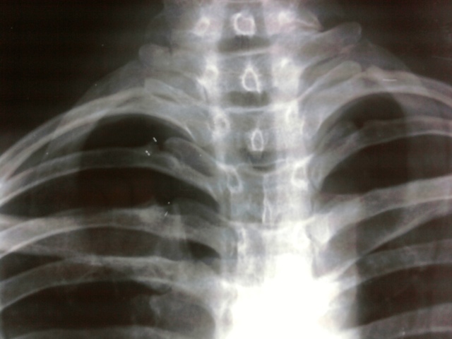
I also recently had xray's of my cervical spine which indicated the following.
1. Loss of normal cervical lordosis. This may be present with spasm.
YOU HAVE LOST THE NATURAL CURVE TO YOUR NECK THIS IS ALSO CALLED HYPOLORDOSIS AND I DO NOT BELIEVE IT IS CAUSED BY SPASM IT WAS CAUSED BY YOUR ACCIDENT.
2. There is old dens fracture present
THE DENS IS AN UPWARD PROJECTION FROM THE SECONG CERVICAL VERTEBRA THAT IS SORT OF LIKE A TOOTH LIKE PROJECTION FROM C2 UPWARDS TO THE C1 VERTBRAE. THIS MEANS YOU ACTUALLY BROKE YOUR NECK IN THIS ACCIDENT. OLD MEANING IT HAS SHOWN SIGNS OF HEALING MY CONCERN HERE IS RADIOLOGIST USUALLY COMMENT "WELL HEALED" FRACTURE. I ACTUALLY WOULD CALL THE RADIOLOGIST AND ASK HIM TO COMMENT FURTHER ON THIS FRACTURE IS IT WELL HEALED OR NOT.
3. Mild degenerative change involving the left neuroforamen at the C5-6 level.
YOU HAVE DEGENERATIVE CHANGES WHERE YOUR SPINAL NERVES EMIT FROM BETWEEN VERTEBRA C5 AND C6 ON THE LEFT SIDE THIS MAY OR MAY NOT BE IRRITATING THE NERVES IN YOUR NECK.
After these xrays I had a cervical MRI done. Which came back normal. So could you please break this down for me and explain what exactly is wrong with me according to the MRI/Xrays.
IF I WERE TREATING YOU I WOULD CALL THE RADIOLOGIST AND ASK HIM IF HE HAD AN OPPORTUNITY TO READ THE X RAYS AND THEN ASK FOR A CLARIFICATION OF "NORMAL"
I am also curious how my cervical xrays could say one thing and my MRI be normal, and if I was diagnosed in 2003 with a C1 fracture but have an old dens fracture present? Isn't the dens considered the C2? Also any advice on treatment and or pain management.
AGAIN I WOULD ASK FOR CLARIFICATION AS YOU ARE CORRECT WITH YOUR ANATOMICAL SITES OF C1 & C2. IT NEEDS TO BE DETERMINED IF YOU IN FACT HAVE TWO SEPARATE FRACTURES. MY COMPLIMENTS ON PICKING UP ON THAT ANATOMICAL FACT. WE NEED A RE-READ WITH THE RADIOLOGIST BEING PRIVY TO ALL REPORTS MRIS AND YOUR HISTORY.
I thank you so much for your time regarding these issues.
PLEASE BE AWARE I OPINE THAT YOU SHOULD HAVE A CHIROPRACTIC WORK UP BUT MUST BE EVER CAREFUL THAT YOU UNDERSTAND THE RISKS OF CERVICAL ADJUSTMENT IN A TRAUMATIC CASE SUCH AS YOURS. YU ABSOLUTELY WILL HAVE TO BE ADJUSTED BY A CHIROPRACTOR THAT IS CONFIDENT IN HIS ABILITY TO ADJUST YOU GIVEN YOUR HISTORY.
THIS IS FOR INFORMATIONAL PURPOSES ONLY AND CANNOT BE TAKEN AS MEDICAL/CHIROPRACTIC ADVICE. THAT DECISION CAN ONL BE MADE ONCE YOU HAVE HAD A PHYSICAL EXAM BY A COMPETENT CHIROPRACTOR.
I sincerely hope this information helps you in your medical decision making.
Dr John Q Quackenbush