QuestionIn December 2008 I lifted a heavy box incorrectly at work. I had immediate pain in low back left hip and down left leg. I'm 38 female I have had two pregnancies full term 18/16 years ago. I work in law enforcement. I wear a duty belt. My job requires me to frequently have to run in boots, generally responding from a rested, seated position. Since the injury I have not been released back to work. Primary GP diagnosed a disc problem had me walk everyday PT X 8 and he prescribed flexeril for spasms. The symptoms of pain tingling vibrations were maddening. I saw a neurologist who gave me Cymbalta for "nerve pain" NSAID for swelling. A Nuero Surgeon looked at my MRI said no disc problem, and he believes I have sacroiliitis. Almost 8 months still have all symptoms of pain numbness tingling lack of mobility. I have had one epidurhal that did not help, it caused pain and I had one SI joint injection that helped for two weeks and two days. All the symptoms have returned. It seems that stretching helps heat and ice help but ultimately I still have localized pain in my SI joint and I can't work. I have been at the mercy of work comp so I can not just go to any doctor I choose. I am at a loss as to what to do for my best health interest. My employer has sent me information that I will see a Qualified Medical Examiner to decide my case. I feel I haven't been properly diagnosed. I will be seeing an orthopedic surgeon. Is there some test I can ask for to determine an actual diagnosis? Does sacroliilitis heal? Can I expect to get better and go back to work? Walking and sitting are painful, over using the hip joint gives me a fever and the muscle spasms are intense. What would you suggest I do to over come this injury? My GP said he thinks I should look into a different line of work? Thank you for your time. It is much appreciated. Teresa
AnswerDear Teresa,
I agree with your neurosurgeon...this definitely sounds like a classic presentation of sacroillitis...I see it often in my practice. The Sacroiliac Joint (or SI Joint,) is the joint between the sacrum and the ilium (one of the bones on the pelvis) The movement in this joint is very little, unlike the movement available in your shoulder. Most commonly, Sacroiliac Pain occurs from dysfunction- either stresses on the joint or too much movement (hypermobility.) This joint has a high concentration of pain receptors due to the large ligaments that stabilize the joint and when inflamed can create significant pain. The fact that your SI joint injections helped to reduce pain is further proof of the problem, but this is likely a structural problem which needs to be addressed in that manner vs. a chemical problem that injections will cure. If the sacrum is not addressed in relationship to the ilium, poor biomechanical relationships and movement patterns will remain which will create chronic pain. Let me explain this further.
The Sacroiliac Joint (SIJ) is kind of L shaped front to back. The surface of one side of the joint (ilial) has a little bulge. The surface of the other side (sacral) has an indentation. This keeps the joint centered. The joint goes out when you are leaning forward to do something and you fail to support the front of your pelvis with you abdominal muscles. The front of the pelvis rocks down (rotating anteriorly or forward) on the front part of the SIJ (the S1 segment) and the back part of the pelvis rotates up and out on the back of the sacrum at the S3 segment. Because the S1 and S3 segments angle in different directions, and the most pressure is at S3, the pelvis slips up and out there and goes a little out of joint (subluxates). This rotation causes the ilial bulge to rise up out of the sacral indentation and flares (spreads) the pelvis. I adjust the sacrum in my clinic due to these issues almost every day...it is one of the most common things I see in practice relating to back pain.
Stresses to the SI Joint can occur from the following activities: persistent standing on one leg, falling on your 揵utt?bone, swinging a golf club, lifting something, or even bending over. If the joint is hypermobile, pain occurs anytime the joint is displaced. This occurs more commonly in females due to their joint structure, hormonal changes, and childbirth strains.
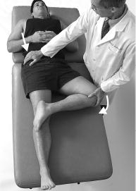
Pain is experienced not only at the joint, but also in the muscles around the SI joint. Pain can also be present in the back of the thigh, in the lower abdomen, or in the groin. Coughing can increase symptoms at the SI joint. Diagnosis of this dysfunction is difficult to see on x-ray because there is very little movement at the joint, unless there is a measurable imbalance of the leg lengths or sacral base and it is often confused with disc injury or sciatica. Physicians often confirm a diagnosis with examination and x-ray analysis, however, specific orhtopedic tests designed to stress the SI joint are the key to making the diagnosis...these are often not performed in the medical setting
Chiropractic and physical rehabilitation is very successful in treating SI Joint Dysfunction. Pain and muscle spasm is addressed with modalities such as ultrasound, electrical stimulation, massage, and heat. Gentle joint adjustments are performed. It抯 key to perform specific stretches on a regular basis. Also, if the abdominals are strengthened properly, they will support the trunk and pelvis. Other strengthening exercises may be prescribed if there are any muscle imbalances present. Lastly, good posture is emphasized.
How Common is SI joint Dysfunction?
In 1992 at the First World Congress on the Sacroiliac Joint Dr. Joseph Shaw of the Topeka Back and Neck Pain Clinic reported that in a series of 1000 consecutive patients he examined for low back pain (LBP) and SIJ he found that 98% had an SIJ problem. When he addressed that problem his surgical incidence for herniated disks dropped to 0.2%.
He stated that he had seen 2000 cases with still the same stats. The problem with Shaw's stats is that they are so good that they seem outrageous. Don't get me wrong, because I believe he is right on the money. The problem is that the 'experts' that are reporting low percentages of SIJD simply cannot believe his results and will not examine the evidence, but if they had done what he did they would have found what he found.
Here is the mind blower. In 1982 the American Academy of Orthopaedic Surgeons met in Toronto specifically to address LBP. They established criteria for testing and for the interpretation of those tests. They assumed that the SIJ was so strong as to be immune to injury through minor trauma and paid scant attention to it. They also reported that 'in spite of thorough examination they could establish a firm diagnosis less than 15% of the time...this is because they aren't examining the joint properly with functional orthopedic tests...chiropractors do this all the time.
What they did not seem to realize is that when you use their recommended tests and interpret those test in the recommended manner that you will be compelled to miss the diagnosis over 85% of the time! It's not that they are not an intelligent group, but they just have not considered all of the evidence. Boorstin once commented that "The chief obstacle to discovery is the illusion of knowledge." People are reluctant to learn what they think they already know. This is willful ignorance. Instruction is of scant value. You must instill some doubt to stimulate investigation....hence I would definitely suggest that you get to a chiropractic physician for management of this issue.
References:
1. DonTigny, Richard. 揗echanics and Treatment of the Sacroiliac Joint,?The Journal of Manual and Manipulative Therapy. Vol.1, No. 1, 1993, pp.3-12.
2. Paris, Stanley V. Introduction to Spinal Evaluation and Manipulation. University of St. Augustine for Health Sciences, St, Augustine, FL, 1997.
3. Slipman et al. "Pain Management Studies Probe Complexities of Sacroiliac Joint Syndrome", Biomechanics, April 2000.
4. Pain Medicine and Management By Mark S. Wallace & Peter Staats. McGraw Publishing 2004.
Now with that being said, lets look at some more current scientific literature on the subject...below is a synopsis of a recent published paper on SI joint pain that I reviewed...as you will see this under diagnosis and improper management of this remains problematic in the medical community.
The Role of Sacroiliac Joint Dysfunction in the Genesis of Low Back Pain:
The obvious is not always right
Archives of Orthopaedic and Trauma Surgery: December 2007 [e-pub]
Authors: Natan Weksler, Gad J. Velan, Michael Semionov, Boris Gurevitch, Moti Klein, Vsevolod Rozentsveig and Tzvia Rudich
FROM ABSTRACT:
Background context: It is a common practice to the link low back pain with protruding disc even when neurological signs are absent. Because pain caused by sacroiliac joint dysfunction can mimic discogenic or radicular low back pain, we assumed that the diagnosis of sacroiliac joint dysfunction is frequently overlooked.
Purpose: To assess the incidence of sacroiliac joint dysfunction in patients with low back pain and positive disc findings on CT scan or MRI, but without claudication or objective neurological deficits.
Methods: Fifty patients with low back pain and disc herniation, without claudication or neurological abnormalities such as decreased motor strength, sensory alterations or sphincter incontinence and with positive pain provocation tests for sacroiliac joint dysfunction were submitted to fluoroscopic diagnostic sacroiliac joint infiltration.
Results: The mean baseline VAS pain score was 7.8. Thirty minutes after infiltration [of the SI joint with anesthesia], the mean VAS score was 1.3. Forty-six patients [out of 50] had a VAS score ranging from 0 to 3, eight weeks after the fluoroscopic guided infiltration.
Conclusions: Sacroiliac joint dysfunction should be considered strongly in the differential diagnosis of low back pain in this group of patients.
THESE AUTHORS ALSO NOTE:
揕ow back pain is second only to common cold as a cause of primary care office visits in the USA.?
揂pproximately 90% of adults have experienced back pain at some point of time in their lives.?br>
Low back pain is responsible for direct care expenditures ranging from $5 billion to more than $20 billion annually and as much as $50 billion per year in indirect costs. Medical residents are not usually taught to consider sacroiliac joint dysfunction as a cause for low back pain.
This study used 200 patients with low back pain who were prior treatment failures from NSAID drugs, physical therapy and intramuscular injections. They were fluoroscopic guided injected with a combination of anesthetic and steroid into their SI joints.
DISCUSSION:
揅hronic persistent low back pain is commonly linked with positive disc findings on CT or MRI imaging. However, these imaging techniques are not always helpful, because they have a poor degree of correlation with clinical signs.? 揑t is not rare to have positive disc findings in asymptomatic patients.?br>
揘early 25% of asymptomatic individuals below the age of 60 years and 33% of older patients have evidence of disc herniation on MRI scans? 揟he diagnosis accuracy of the tissue origin of chronic low back pain and referred lower extremity symptoms based on clinical criteria are about 19?4%.?[In medical practice] Estimated prevalence of SI joints causing low back pain is 13 to 30%.
揟he SIJ has a rich nociceptive innervation.?Its anterior portion is innervated by the posterior rami of the L2朣2 roots, and its posterior aspect is innervated by the posterior rami of L4朣3. [IMPORTANT]
The piriformis muscle is located close to the SIJ, originating at the anterior aspect of the sacrum and inserting into the greater trochanter of the femur. SIJ problems can cause piriformis spasm and provoke sciatic irritation, with 損ain radiating to the buttock, the posterior calf, and to the anterior and lateral calf and foot mimicking radiculopathy.?[IMPORTANT]
揟he current gold standard for the diagnosis of the SIJ syndrome is fluoroscopically guided infiltration of local anesthesia leading to at least an 80% reduction in VAS scores.?However, three physical tests showed good correlation with the diagnosis of SIJ dysfunction: SIJ compression test (94% agreement), the thigh thrust test (90%) agreement, Yeoman抯 test (88%) agreement. [IMPORTANT]
Studies have 揷oncluded that manipulations appear to be successful in many patients suffering from SIJ dysfunction?
CONCLUSIONS:
1) The incidence of SIJ dysfunction in patients with low back pain and discopathy on CT or MRI scans and without neurological deficits appears to be higher than previously described.
2) Pain in SIJ dysfunction can radiate towards the calf and foot mimicking radicular pain.
3) 揚hysicians seeing patients with low back pain should have a high index of suspicion for SIJ dysfunction, especially in the absence of neurological deficits.?
KEY POINTS FROM SUNCOAST HEALTHCARE PROFESSIONALS
1) Because pain caused by sacroiliac joint dysfunction can mimic discogenic or radicular low back pain, the diagnosis of sacroiliac joint dysfunction is frequently overlooked. However, chiropractic physicians are specifically trained to evaluate SIJ function along with lumbar spine function. [IMPORTANT]
2) 揕ow back pain is second only to common cold as a cause of primary care office visits in the USA.?
3) 揂pproximately 90% of adults have experienced back pain at some point of time in their lives.?br>
4) Medical residents are not usually taught to consider sacroiliac joint dysfunction as a cause for low back pain. [IMPORTANT]
5) 揅hronic persistent low back pain is commonly linked with positive disc findings on CT or MRI imaging. However, these imaging techniques are not always helpful, because they have a poor degree of correlation with clinical signs.?br>
6) 揘early 25% of asymptomatic individuals below the age of 60 years and 33% of older patients have evidence of disc herniation on MRI scans?br>
7) Estimated prevalence of SI joints causing low back pain is 13 to 30%.
8) The SI joint has a rich nociceptive innervation by the posterior rami of the L2朣3 roots. [IMPORTANT]
9) The piriformis muscle is located close to the SIJ, originating at the anterior aspect of the sacrum and inserting into the greater trochanter of the femur. SIJ problems can cause piriformis spasm and provoke sciatic irritation, with 損ain radiating to the buttock, the posterior calf, and to the anterior and lateral calf and foot mimicking radiculopathy.?[IMPORTANT]
10) 揟he current gold standard for the diagnosis of the SIJ syndrome is fluoroscopically guided infiltration of local anesthesia leading to at least an 80% reduction in VAS scores.?
11) Three physical tests showed good correlation with the diagnosis of SIJ dysfunction: SIJ compression test (94% agreement), the thigh thrust test (90%) agreement, Yeoman抯 test (88%) agreement. [IMPORTANT]
12) Manipulation is successful in many patients suffering from SIJ dysfunction. [IMPORTANT]
13) The incidence of SIJ dysfunction in patients with low back pain and discopathy on CT or MRI scans and without neurological deficits appears to be higher than previously described.
14) Pain in SIJ dysfunction can radiate towards the calf and foot mimicking radicular pain. [IMPORTANT]
15) 揚hysicians seeing patients with low back pain should have a high index of suspicion for SIJ dysfunction, especially in the absence of neurological deficits.?br>
16) Sacroiliac joint dysfunction should be considered strongly in the differential diagnosis of low back pain in this group of patients.
Hope this helps Teresa.
Respectfully,
Dr. J. Shawn Leatherman
www.suncoastehalthcare.net