The synovial membrane is an important structure that lines all the synovial joints in the human body. This Buzzle article highlights details regarding the structure, function, and diseases affecting the synovial membrane.
The synovial membrane is a tissue layer that forms a bordering line along the synovial joints. This membrane is made up of a soft tissue that lines the non-cartilaginous surfaces within joints, which have cavities (synovial joints). The word 'synovium' comes from the Latin word that means 'with egg', because the synovial fluid that is present in the joints resembles an egg white. This is an important membrane that acts as a lubricating agent, thus aiding free movement of these joints.
Structure of the Synovial Membrane
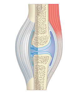
Although the structure of the synovium can vary, it is generally made up of two layers. The outer layer is also known as
subintima. It can be of almost any type, i.e., made up of fibrous, fatty, or loosely areolar tissue. The inner layer is also known as
intima. It consists of a sheet of cells, the thickness of which is lesser than that of a paper. The subintima is loose and the intima sits on a pliable membrane. This membrane, together with the cells of the intima, acts as an inner tube, which seals off the synovial fluid from the surrounding tissue. This is a protective reflex, which helps to prevent the joint from getting squeezed when subject to an impact. The intimal cells are classified into two types -
fibroblasts and
macrophages.
Fibroblasts and Macrophages
Fibroblasts are responsible for constructing long-chain sugar polymers known as hyaluronan. This gives the synovial fluid its ropy consistency. This along with a molecule called lubricin helps to lubricate the joint surfaces. The macrophages are responsible for ingesting foreign molecules that are harmful. The surface of the synovium may be flat or covered with finger-like projections known as villi. These protrusions help to allow the soft tissue to change shape as the joints move. The blood supply is carried out by a dense net of blood vessels that are present just beneath the intima. These help to provide nutrients to the synovium and the avascular cartilage. The synovial membrane and periosteum are also very close to each other and at times, some areas of the adjoining cartilage need to obtain nutrients indirectly, and they may do so by diffusion through the cartilage.
Function
Contrary to common belief, the space in which the synovial fluid is lodged is not very large. Thus, the membrane has a variety of functions, the most important of which is to provide a plane of separation or disconnection between solid tissues so that the movement can occur smoothly without any friction. The synovial membrane also helps to act as a packing that can change shape in the way that is needed for easy movements. This is the reason it needs to be so flexible. The membrane also has the job of controlling the volume of fluid that is present in the synovial cavity so that it is just enough to allow the solid components to move freely over each other. Any sudden change in this volume could result in various diseases and disorders.
Pathologies
Synovial Membrane Disorder
Presentation of Disease
Rheumatoid arthritis
Excess of synovial cell proliferation, edema and basically synovial membrane inflammation
Scleroderma
Superficial fibrin deposits
Hemochromatosis
Excess deposition of iron in synovial lining cells
Tuberculous arthritis
Caseating granulomas along with giant cells
Alkaptonuria
Shards of pigmented cartilage
Pigmented villonodular synovitis
Hypertrophy of villi
Gout
Deposition of crystals made of monosodium urate
Neoplasms
Malignant cells in synovium
This structure plays a very important role in ensuring that there is a smooth functioning of all the synovial joints in the body.