Fractured neck of femur patients take up significant numbers of beds in hospitals in all the developed nations, as elderly people develop low bone density, especially women after the menopause. Less commonly stress fractures can occur in anyone who puts greatly increased stresses through their hips such as running athletes and soldiers who are in a much younger age group. Trauma such as a fall onto the side of the hip can cause this fracture in almost any age group as can pathological conditions in the area secondary to tumours.
Orthopaedic practitioners have long recognised the importance of promptly restoring the anatomical alignment of the femoral neck fracture so that avascular necrosis (AVN) of the femoral head could be avoided. In AVN the blood supply is lost to the head of the femur, causing death of the bone and the head then degenerates and collapses, necessitating replacement operation. Immobilisation in a hip spica was initially used, with internal fixation developed in the 1930s by Smith-Petersen. Later developments such as the Richards Screw Plate use a form of sliding fixation which allows surgical impaction of the fracture site.
Compressive and shear stresses pass across the femoral necks when we do normal things such as walk but they can be greatly increased by involvement in sports such as jumping, high athletic performance and jogging. The bodyweight can be amplified five or six times across the hip in fairly standard activities such as stair climbing, let alone sport. The groin, the lateral hip and the anterior thigh are the typical areas of presentation of hip related pain from many pathological changes as well as stress fractures, which may worsen to complete fractures with or without displacement with its risks and complications.
In younger healthy people who exert abnormally high demands on normal bone the bone structures can fail mechanically due to the excessive stresses imposed on them. In older people, especially post menopausal women, normal stresses are imposed on bone which is not able to cope with them, bone with pathological changes due to insufficiency of the bone from osteoporosis or other metabolic abnormality. Oestrogen maintains the turnover and health of bone strength and without it bones become more brittle, either in older women or female athletes in high intensity training.
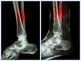
An athlete with insidious onset of hip pain after an increase in training or new activity should be considered as potentially having a stress fracture, typically better with resting and worse with the sport. Bone scanning can be used to investigate this as its sensitivity is higher than standard x-rays. However, it is much more common for a femoral neck fracture to present after a fall in an elderly person. This fracture presents with a short leg, the inability to bear weight on the hip, a laterally rotated hip and pain in the lateral hip or groin.
There is a ten to fifteen percent chance of transverse femoral neck fractures displacing with the consequent problems of avascular necrosis. Typically these fractures require surgical management and the orthopaedic specialist must choose the correct approach. The anatomical position of the fracture dictates the subsequent management, being either the replacement of the whole joint or internal fixation of the fracture. Sub-capital fractures, just below the femoral head, are likely to interfere with the circulation and in these cases joint replacement or Thompson hemiarthroplasty are preferred.
If the fracture is not displaced and is under compression due to the nature of the injury then conservative management is indicated and surgery unnecessary. If the fracture is under tension it is unstable and has the potential to displace and requires internal fixation with one of the many fracture fixation devices. This applies to lower femoral neck fractures, trochanteric and sub-trochanteric fractures. Fractures are fixed as soon as possible after the injury, allowing for the medical condition of the patient.
Once the fracture is replaced or fixed the patient is allowed 24 hours to recover medically then the physiotherapist and an assistant will check the operative instructions, review the patient's observations and get the patient up weight bearing with a frame or crutches.