To understand spinal stenosis, it helps to understand the anatomy of your spine. The spine is a column of connected bones called vertebrae. There are 24 vertebrae in the spine, plus the sacrum and tailbone (coccyx). Most adults have 7 vertebrae in the neck (the cervical vertebrae), 12 from the shoulders to the waist (the thoracic vertebrae), and 5 in the lower back (the lumbar vertebrae). The sacrum is made up of 5 vertebrae between the hipbones that are fused into one bone. The coccyx is made up of small fused bones at the tail end of the spine.
At the back (posterior) of each vertebra, you have the lamina, a bony plate that protects your spinal canal and spinal cord. Your vertebrae also have several bony tabs that are called processes; those processes are attachment points for muscles and ligaments. Vertebrae are connected by ligaments, which keep the vertebrae in their proper place.
The ligamentum flavum is a particularly important ligament. Not only does it help stabilize your spine, it also protects your spinal cord and nerve roots. Plus, the ligamentum flavum is the strongest ligament in your spine.
The ligamentum flavum is a dynamic structure, which means that it adapts its shape as you move your body. When you're sitting down and leaning forward, the ligamentum flavum is stretched out; that gives your spinal canal more room for the spinal nerves. When you stand up and lean back, though, the ligamentum flavum becomes shorter and thicker; that means there's less room for the spinal nerves. (This dynamic capability helps explain why people with spinal stenosis find that sitting down feels better than standing or walking. You can learn more about the causes of spinal stenosis in the article Causes of Spinal Stenosis.)
In between each vertebra are tough fibrous shock-absorbing pads called the intervertebral discs. Each disc is made up of a tire-like outer band (annulus fibrosus) and a gel-like inner substance (nucleus pulposus).
Nerves are also an important part of your spinal anatomy—after all, they're what sends messages from your brain to the rest of your body. The spinal cord, the thick bundle of nerves that extends downward from the brain, passes through a ring in each vertebra. Those rings line up into a channel called the spinal canal. Between each vertebra, two nerves branch out of the spinal cord (one to the right and one to the left). Those nerves exit the spine through openings called the foramen and travel to all parts of your body.
Nerve Structures
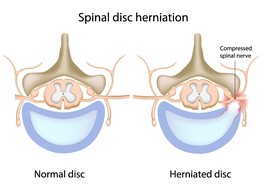
Normally, the spinal channel is wide enough for the spinal cord, and the foramen are wide enough for the nerve roots. But either or both can become narrowed—that'd be the spinal stenosis—and lead to pain.
Copyright © www.orthopaedics.win Bone Health All Rights Reserved