Compressive Forces
A compression fracture results from tension created by external force (e.g. trauma). The compressive force can cause one or more vertebrae to crush, burst, or wedge. The fracture may 'compress' the spinal cord or nerve roots resulting in neurologic deficit. These fractures cause acute pain.
Osteoporosis
The risk for compression fracture increases with age. Osteoporosis, a bone weakening disease greatly increases the risk for compression fracture. Trauma (e.g. falling) or simply bending may cause compression fracture in an osteoporotic spine. Over time, thoracic compression fractures can create a kyphosis (e.g. humpback) accompanied by disabling pain and neural compromise.
Nerve Injury
The degree of neurologic injury is usually due to the amount of force present at the time of injury and the amount of compromise of the spinal canal. If the fracture bursts, bony fragments can be forced into the spinal canal causing loss of spinal cord function. This may cause loss of strength, sensation, or reflexes below the level of injury. In an incomplete spinal cord injury only partial paralysis or reflex loss is seen. With mild fractures only transient symptoms may be present or no neurologic injury may be present.
Diagnosis
When spinal fracture is suspected, do not move the patient. They should be kept lying flat and transported in a flat position. Movement or sitting may increase the risk for neurologic injury. Compression fractures require immediate medical care and the patient should be transported to an emergency room.
X-rays and a CAT Scan are used to make the diagnosis. An MRI may be ordered to assess soft tissue damage, bleeding, or ligament disruption. These tests allow the treating physician to make a determination as to the level of the fracture, the type, and if there is spinal canal compromise. All of these factors enter into the treatment decision process.
A physical examination should be performed to document spinal deformity and tenderness at the fracture level. A neurologic exam includes testing muscle strength, sensation and reflexes of the lower extremities, as well as, bowel and bladder sphincter control.
If osteoporosis is suspected, a Bone Mineral Density (BMD) test may be ordered. Depending on the patient's condition, laboratory tests including complete blood count, urinalysis, and thyroid function may be required.
Treatment
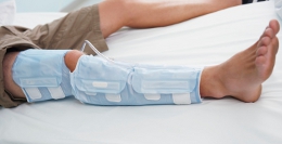
Kyphoplasty and Percutaneous Vertebroplasty (PV) are two types of surgical procedures used to treat compression fractures. These procedures have benefitted many patients with osteoporosis. A spine specialist determines if this procedure will benefit the patient. The spine specialist may recommend a brace or corset to immobilize the affected area for a period of time. Some patients simply cannot tolerate wearing an orthotic because of pain. Compression fractures occurring in the cervical spine may be immobilized using a Philadelphia Collar or other cervical orthotic.
Analgesics, anti-inflammatories, and/or muscle relaxants may be prescribed.
Recovery
The recovery time is dependent on neurologic deficit. Most patients without neurologic deficit can make a near complete recovery and return to most activities. Physical therapy may be prescribed to build strength, flexibility, and increase range of motion. Additionally, the therapist provides the patient with a customized home exercise program.
Copyright © www.orthopaedics.win Bone Health All Rights Reserved