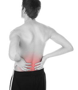
Diagnosing the form of spinal stenosis that you have may not be as simple as it seems seeing as the various forms of spinal stenosis will produce similar symptoms. The most common symptoms for lumbar spinal stenosis are leg pain, often referred to as sciatica as well as some low back pain, leg numbness and tingling, and having limitations in walking.
Although leg pain from walking can be one of the symptoms of spinal stenosis, it may also be an indication of arterial circulatory insufficiency. Rest will help to ease the leg pain from either condition but a tell-tale sign of spinal stenosis is that the patient will usually have to sit down for a few minutes to feel relief from the pain. If the patient affected by vascular claudication simply stops walking the pain will go away.
Generally speaking the symptoms of spinal stenosis develop over the course of several years, and occasionally these symptoms may become acute. With most spinal stenosis victims their leg pain will worsen the longer then stand or walk. One of the ways to relieve the leg pain and other symptoms associated with spinal stenosis is flexing forward or sitting, but most often the pain recurs when the patient resumes an upright posture. Numbness and tingling often accompany the pain of spinal stenosis, although true weakness can be a rare symptom as well. If you see an older person leaning over the handle of their shopping cart while making short stumbling steps this is often a sign that that person is afflicted with spinal stenosis.
A general quick reference for spinal stenosis is often characterized as follows:
- Spinal stenosis develops slowly over time.
- Most often the pain comes and goes and is not acute.
- Walking and standing upright often brings on the symptoms.
- Painful symptoms can be relieved by rest, either sitting or lying down. Flexing yourself forward also helps to relieve your symptoms.
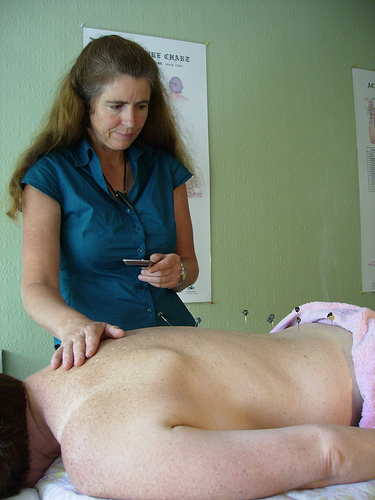
Either a MRI (Magnetic resonance imaging) or a CT (Computed tomography) with myelogram (uses an x-ray dye in the spinal sack fluid) imaging studies are used to diagnose spinal stenosis. Unless the CT scan with is preformed with very fine segmental scan slices it is often of little value.
Spinal stenosis has a dynamic effect on nerve compression. This in short means that when bearing weight on the affected area there will be symptoms and when there is no pressure on the area there will be no symptoms.
Because of this changing compression, physical examinations will generally not show and problems or motor weakness. There are some recent scanning methods that allow for studying the effects of spinal loading on the upright body position.
Although foraminal stenosis can be pinpointed by a CT or MRI scan, selective nerve blocking can also be preformed. This is done by injecting the suspected nerve with a small amount of local anesthetic. If successful, after the injection there will be a remission of the spinal stenosis symptoms. Once clinically diagnosed the patient will be more informed and can make a better decision on the possibility of surgery.